1. LON-CAPA Logo
  2. Help
  3. Log In


tRNAPhe

from yeast


To highlight described properties click the boxes . Move the molecule anytime with the mouse - if the view is messed up, clicking the large boxes will restore a convenient view.

Transfer RNAs show a multitude of structural features apart from the 'ordinary' helical duplex with strict basepairing as in DNA. In contrast to linear nucleic acids whose main purpose is to contain genetic information tRNA has rather a compact structure adopted to its role in protein synthesis. If viewed more abstractly as the backbone of its RNA chain some double helical stretches are seen. The other regions are associated with chemically modified bases (14 out of 76) and/or mispaired or unpaired bases.

The following lists all nucleosides found in tRNAPhe (the models shown here have different conformations from their occurences in tRNA):
adenosine
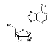
Adenosine
(A, 5,9,13,14,21,23,29,31,35,36,38,39,44,62,64,66,67,73,76)

in tRNA: show - hide       reset
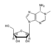
methyladenosine
1-methyladenosine (1MA, 58)

in tRNA: show - hide       reset
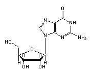
guanosine
Guanosine
(G, 1,3,4,15,18-20,22,24,30,42,43,45,51,53,57,65,71)

in tRNA: show - hide       reset
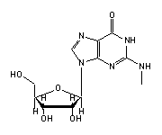
2m-guanosine
N2-methylguanosine (2MG, 10)

in tRNA: show - hide       reset

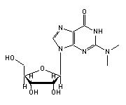
dimethylguanosine
N2-dimethylguanosine (M2G, 26)

in tRNA: show - hide       reset

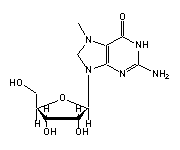
7-methylguanosine
7-methylguanosine (7MG, 46)

in tRNA: show - hide       reset

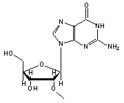
O-methylguanosine
O2'-methylguanosine (OMG, 34)

in tRNA: show - hide       reset
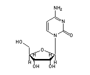
cytidine
Cytidine
(C, 2,11,13,25,27,28,48,56,60,61,63,70,72,74,75)

in tRNA: show - hide       reset
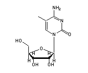
5-methylcytidine
5-methylcytidine (5MC, 40,49)

in tRNA: show - hide       reset

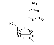
O-methylcytidine
O2'-methylcytidine (OMC, 32)

in tRNA: show - hide       reset
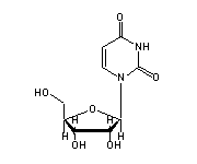
uridine
Uridine
(U, 6-8,12,33,41,49,50,52,59,68,69)

in tRNA: show - hide       reset
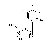
methyluridine
5-methyluridine (5MU, 54)

in tRNA: show - hide       reset

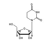
dihydrouridine
5,6-dihydrouridine (H2U, 16,17)

in tRNA: show - hide       reset

pseudouridine
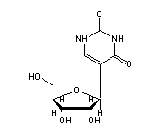
Pseudouridine (PSU, 39,55)

in tRNA: show - hide       reset

wybutosine
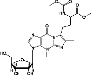
Wybutosine (YG, 37)

in tRNA: show - hide       reset

Basepairings in tRNAPhe are:

  G 1 - C 72
  C 2 - G 71
  G 3 - C 70
  G 4 - U 69 : non-Watson-Crick
  A 5 - U 68
  U 6 - A 67
  U 7 - A 66
  U 8 - A 14 - A 21 : non-W-C/triple pair
  A 9 - U 12 - A 23 : triple pair
  2MG 10 - C 25 - G 45 : triple pair
  C 11 - G 24 - G 45 : triple pair
  C 13 - G 22 - 7MG 46 : triple pair
  G 15 - C 48 : non-Watson-Crick
  H2U 16 - U 59 : non-Watson-Crick
  G 18 - PSU 55 : non-Watson-Crick
  G 19 - C 56
  M2G 26 - A 44 : non-Watson-Crick
  C 27 - G 43
  C 28 - G 42
  A 29 - U 41
  G 30 - 5MC 40
  A 31 - PSU 39 : non-Watson-Crick
  OMC 32 - A 38 : non-Watson-Crick
  5MC 49 - G 65
  U 50 - A 64
  G 51 - C 63
  U 52 - A 62
  G 53 - C 61
  5MU 54 - 1MA 58 : non-Watson-Crick

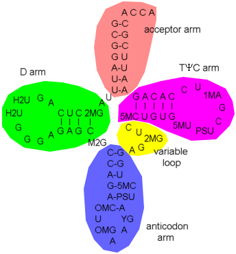
  nucleotides from acceptor to anticodon

Unpaired bases

 in acceptor arm (A73, C74, C75, A76)
 in D arm (H2U17,G20)
 in anticodon arm (U33, YG37)
 in anticodon (OMG34, A35, A36)
 in variable loop (U47)
 in T psi C arm (G57, C60)

The 'arm' designation originates from the time when only the secondary structure was known ('cloverleaf') and is somewhat misleading in the tertiary structure  .
cloverleaf

The structures marked red and dark green in the left frame are magnesium ions partly coordinated with water molecules. They stabilize the tRNA once it is folded to its final form. One magnesium in the variable loop is completely (tetrahedrally) coordinated with water (shown as red spheres here)  . The waters are hydrogen bridged to the phosphate backbone of U8 to U12  . Another magnesium surrounded by six waters is situated in the anticodon arm and held by G24, C25 and 5MC40 - G42  . Some more tightly bound water molecules are involved in hydrogen bridges  . Other magnesiums are partly coordinated to the backbone phosphate groups

by G19
by G20, A21

The backbone of the tRNA is partly stabilized by water molecules as shown here for the T psi C arm (G51 - U59)  . The water molecules bind to posphate oxygens to bridge two nucleotides each  .
Some twin water molecules bridge the ribose 2' hydroxyl group to a phosphate oxygen of the next nucleotide  .

The most rigid part of the tRNA is the central domain formed by the D arm and the T psi C arm (which are folded tightly together) as judged from the temperature factor of the X-ray data (blue = rigid, red = movable).


Literature:
H Shi & PB Moore, The crystal structure of yeast phenylalanine tRNA at 1.93 Å resolution: A classic structure revisited, RNA 6 (2000) 1091-1105
L Jovine et al, The crystal structure of yeast phenylalanine tRNA at 2.0 Å resolution: Cleavage by Mg2+ in 15-year old crystals, J. Mol. Biol. 301 (2000) 401-414


10-01 - R Bergmann