
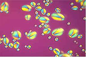
Light waves do usually radiate into all directions. Suitable polarization masks allow the elimination of certain planes of polarization, whereby linear polarized light is gained. It can be completely erased by a second polarization mask that is placed at a right angle to the first one. Such polarization masks that can rotate around their own axis can be placed in the light cone of a microscope. One beneath the condenser and the other above the objective.
 The use of a thus constructed polarization microscope is only sensible,
if preparations with polarizing properties are examined. This is the
case, if the preparation consists of units that are oriented in a
certain way like molecules or atoms (double refracting crystals).
This type of microscope is thus used mainly in mineralogy since
crystals are per definitionem built in a regular pattern. By
the use of polarized light crystal axis and three-dimensional can
pattern be ascertained exactly. The practical application in biology
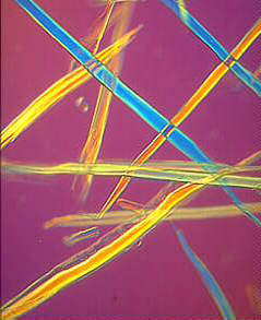
is somewhat limited, but the structure of starch grains for example
or the orientation of cellulose fibrils within the vegetable cell
wall (figure to the right) or the orientation of rod-shaped viruses
(tobacco mosaic virus) can be determined.
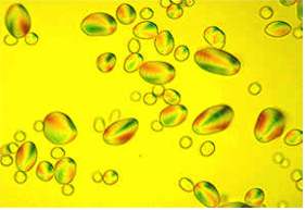
The use of a thus constructed polarization microscope is only sensible,
if preparations with polarizing properties are examined. This is the
case, if the preparation consists of units that are oriented in a
certain way like molecules or atoms (double refracting crystals).
This type of microscope is thus used mainly in mineralogy since
crystals are per definitionem built in a regular pattern. By
the use of polarized light crystal axis and three-dimensional can
pattern be ascertained exactly. The practical application in biology
is somewhat limited, but the structure of starch grains for example
or the orientation of cellulose fibrils within the vegetable cell
wall (figure to the right) or the orientation of rod-shaped viruses
(tobacco mosaic virus) can be determined.
 Based
on polarization microscopy the French physicist G. NOMARSKI developed
the interference contrast
microscope (also called differential interference
contrast, DIC) in Paris during the middle of the 1950s. For this type
of microscopy two additional Wollaston-prisms are needed. A
Wollaston-prism is composed of two cemented calcium fluoride wedges.
A polarized ray of light is split into two beams at the cemented
plane that are vertical towards each other . The first
Wollaston-prism is used in the front focal plane of the condenser,
the second in the back focal length of the objective. The object is
thus passed by two beams that are vertical to each other. The phase
of the beams is shifted according to the thickness and the refraction
characteristics of the object. Optimal interference contrast is
reached at the rims of the object, where the phase of both beams is
shifted differently. It is important, however, how the object is
orientated. A rotating microscope stage is used to analyse the object
in all its orientations. The second Wollaston-prism reunites the
rays. To achieve interference the planes of oscillation have to
coincide. This is done by the polarization mask above the objective.
An interference contrast image looks like a three-dimensional relief.
An inexperienced microscoper could thereby be lead to the
misapprehension of a three-dimensional structure of the preparation.
This is not the case. The tree-dimensional appearance stems from the
fact that the differences in the density of the preparation are
transformed into differences in height in the image. In contrast to
phase-contrast microscopy even relatively thick preparations can be
analyzed.
Based
on polarization microscopy the French physicist G. NOMARSKI developed
the interference contrast
microscope (also called differential interference
contrast, DIC) in Paris during the middle of the 1950s. For this type
of microscopy two additional Wollaston-prisms are needed. A
Wollaston-prism is composed of two cemented calcium fluoride wedges.
A polarized ray of light is split into two beams at the cemented
plane that are vertical towards each other . The first
Wollaston-prism is used in the front focal plane of the condenser,
the second in the back focal length of the objective. The object is
thus passed by two beams that are vertical to each other. The phase
of the beams is shifted according to the thickness and the refraction
characteristics of the object. Optimal interference contrast is
reached at the rims of the object, where the phase of both beams is
shifted differently. It is important, however, how the object is
orientated. A rotating microscope stage is used to analyse the object
in all its orientations. The second Wollaston-prism reunites the
rays. To achieve interference the planes of oscillation have to
coincide. This is done by the polarization mask above the objective.
An interference contrast image looks like a three-dimensional relief.
An inexperienced microscoper could thereby be lead to the
misapprehension of a three-dimensional structure of the preparation.
This is not the case. The tree-dimensional appearance stems from the
fact that the differences in the density of the preparation are
transformed into differences in height in the image. In contrast to
phase-contrast microscopy even relatively thick preparations can be
analyzed.
Besides the light-dark-contrasts, that can be enhanced or diminished by the rotation of the polarizator or by the adjustment of the second Wollaston-prism, colour contrasts can be achieved by the use of a lambda / 4 mask.
The Wollaston prism splits light beams. The points and lines show the polarization direction of the respective beams. (according to a works photography of CARL ZEISS)
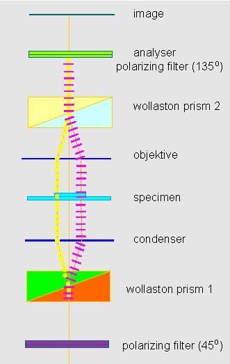
Path of light rays within an interference contrast microscope.
|
|