Secondary dermal tissues are usually found in older shoots and roots, because these are the organs where secondary growth takes place. Only rarely and only in their initial stages do secondary tissues occur in bryophytes, ferns and monocots. They stem from the phellogen, a secondarily developed cambium. The cells of the phellem are called cork cells, they are generated centrifugally, are non-living and have suberized cell walls. The phelloderm consists of cells given off towards the inside of the phellogen, forming the inner part of the periderm. Periderm is the collective term for all three types of tissue.
Cork layers are neither permeable by water nor gas and do only rarely constitute homogeneous structures. The cracked surface structure of the bark that typical for certain species is a visible expression of this.
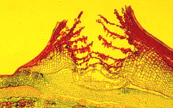
The phellem is often interspersed with groups of parenchyma cells of elongated appearance, called lenticels. These can be observed on the surface of elder (Sambucus niger), a typical specimen of basic botanical courses.
The lenticels serve to permit gas-exchange
between the metabolically active cells below the bark and the
atmosphere. They are not only found in the shoot, but in roots, too.
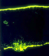
Cork is rarely found within primary tissues, the leaves of
Tabernnaemontana pachysipon are an exception. They have little
islets of cork at the undersurface of their leaves.
The totality of all tissues produced by the phellogen, both primary and secondary ones as well as some other types, is called bark. You could simplify things by saying that all tissue outside the cork cambium is bark.

Lenticel from the bark of the tropical leguminosa
Entada rhedii (Mimosaceae)

Tabernaemontana pachysiphon (Apocynaceae): cross-section through a leaf with a cork cell aggregate at the leaf undersurface. Cuticle and cork cells are stained with the fluorescent dye berberin sulphate.
|
|