
The other vascular tissue is the phloem. Its principal function is the conducting of assimilates and food. It is composed of the sieve elements, of which two types can be distinguished, sieve cells and sieve-tube members. Phloem elements do typically have sieve plates instead of final walls. Sieve-tube members of angiosperms are associated by living companion cells.
The phloem is the principal food-conducting tissue of vascular plants. Its elements are elongated, just like those of the xylem. In contrast to tracheids and wood vessels, mature phloem elements contain a protoplast and sometimes even a nucleus. Phloem elements may be of primary or secondary origin though the early primary phloem, the protophloem, is frequently destroyed during elongation of the resepctive organ.
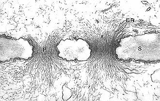
The main conducting elements of the phloem are the sieve elements, of which there are two different types: sieve cells and sieve-tube members. Sieve cells have narrow pores, their sieve areas are quite uniform in structure, and they are distributed evenly on all walls. One of the principal differences between sieve cells and sieve-tube members is the presence of sieve plates in sieve-tube members, that are absent in sieve cells. Sieve cells are the only type of food-conducting cells in most seedless vascular plants and gymnosperms, whereas in angiosperms only sieve-tube members are present. Sieve-tube members occur end-on-end in longitudinal series called sieve tubes. They are in contact via plasmodesmata. Typically, the final walls are interspersed with primary pit areas (groups of plasmodesmata), that later on develop into sieve plates. Sieve tubes in the phloem of angiosperms are flanked by one or several plasma-rich, nucleated companion cells, that do not occur in gymnosperms.
Much less is known about the phylogeny of sieve elements than about that of xylem members. One cause are the cell walls of sieve elements, that are strengthened entirely by cellulose and are therefore not as resistant as lignified ones, another is the transitory working order of the single sieve element. After loss of function the cells are reorganized and lose their typical structural properties. Good fossils are thus rare.
Despite these obstacles it was possible to reveal in comparative plant anatomical studies (of monocots) definite phylogenetic trends (V. I. CHEADLE, 1948, CHEADLE et al., 1941, 1948, K. ESAU et al., 1953, ESAU, K., 1964/69):
- The originally scattered pits are concentrated in 'sieve areas' (accumulations of pores). The diameter of a pore is around 0.1 - 15 µm.
- Specialized sieve areas are concentrated in the final walls.
- A gradual change of orientation of these final walls from very sloping to right angles occurs.
- The change is followed by a stepwise transition from compound to simple sieve plates.
- The activity of the sieve areas in the side walls is reduced. This leads to canalizing of the flow of assimilates in longitudinal direction and the long-distance transport rates are enhanced.
Phloem elements of the roots are normally organized in a more progressive and thus more efficient way than their equivalents in the shoots. Callose is deposited in the sieve plates at regular intervals and can be detected with special reagents. Resorcin blue gives a blue, aniline blue a bright yellow staining. The amount of callose increases with cell age, continuously reducing the diameter of the pores. Sieve elements that have lost their function are blocked by thick plugs of callose. The question, what is cause and what result remains. Are the cells inactivated by the callose plugs or do these form as a result of the loss of function?
 Active
sieve tubes contain huge amounts of so-called
P-proteins (phloem - proteins).
It has often been speculated, whether they have an active part in
assimilate transport. No answer has been found up until now.
Active
sieve tubes contain huge amounts of so-called
P-proteins (phloem - proteins).
It has often been speculated, whether they have an active part in
assimilate transport. No answer has been found up until now.
The sieve tube elements of angiosperms (mono- and dicots), but not those of pteridophytes and gymnosperms are associated with companion cells. In Ginkgo and other gymnosperms their function is taken over by specialized parenchyma cells, that resemble companion cells in structure: the albuminous cells. They work out the contacts between phloem and the surrounding transfusion parenchyma and can also be found as mediators between ray parenchyma cells of the bark and mature sieve cells. Albuminous cells participate in loading and unloading of these cells. The process has been studied in detail in Pinus.
Companion cells and sieve elements are of meristematic origin, their development is similar to that of the xylem. Primary phloem develops by longitudinal division and subsequent elongation of meristematic cells. The cells do often divide unequally. The bigger daughter cell differentiates into a sieve element, the smaller after one or two further longitudinal divisions into two to four companion cells. Consequently several companion cells may belong to one sieve cell. The exact number is usually typical for the respective tissue, but it may even vary within a single plant.
Beside the typical phloem elements, fibres and/or sclereids as well as parenchyma cells that serve as deposits for starch, fat, oil and other nutrients, can belong to the phloem. The deposits contain often tannic acids and resins.
|
|