
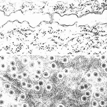
a: Longitudinal section. Plasmodesma at a young cell wall between haustorial cells of dodder (Cuscuta odorata). The endoplasmatic reticulum of the two cells forms a continuous system through the plasmodesmata. Both cells are rich in rough ER.
(Ch. GLOCKENMANN, R. KOLLMANN, 1975) b: Cross-section (top view). Plasmodesmata with callose cylinders in a wall between phloem parenchyma cells in the shoot of Metasequoia glyptostroboides. (Ch. GLOCKMANN, R. KOLLMANN, 1979).