
Components of the cytoplasm in an epidermal cell of Allium cepa. The cells of the inner epidermis of the onion were examined with an UV-microscope at a wave length of 310 nm. This wave length caused no visible changes of the cytoplasm during the period of observation (5-10 min). The higher resolution of this microscope compared to conventional light microscopes allows a clear depiction of organelles. Further elements become visible that are hardly ever observed with a light microscope and are only known from electron and fluorescence microscopy. They are thin elements of the endoplasmatic reticulum (ER) with a diameter of roughly 100 nm and ramified bundles of actin filaments where directed and active movements of organelles take place. The diameter of one of these bundles is 100 nm or less
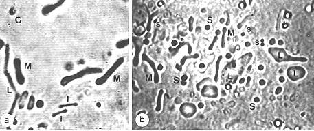
a:M Mitochondrion, l Small, mitochondria-like inclusions, L Leucoplast, G Golgi-apparatus (side view). b: Legend like in a., additionally S and ss: Large and small sphaerosomes (membrane-surrounded vesicles) and particles with unknown function.
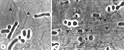
c: Thin elements of the ER are often arranged as a polygonal network (A) in unstreaming plasma that is located at the cell wall. d: In streaming plasma the ER-elements often have the same orientation as the plasma streaming.
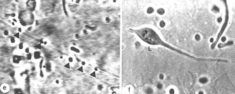
e: Bundles of actin filaments with wandering sphaerosomes. f: L Leucoplast (in top view with process).
(a, b. I. K. LICHTSCHEIDL and W. G. URL, 1987, c - f. I. K. LICHTSCHEIDL, 1987)
© Peter v. Sengbusch - b-online@botanik.uni-hamburg.de