
Specimen: Cells of the inner epidermis of Allium cepa. a and b are examples of digital contrast enhancement by subtraction

a. Unprocessed picture: photo taken with an interference contrast microscope. When studying plasma close to the cell wall the structure of the cell wall is especially interfering. b. the unprocessed picture was taken 10 sec after picture a with another focal plane. Subsequently a was subtracted from b. In the resulting picture show the plasmatic components with enough contrast. A polygonal pattern formed from ER-elements becomes clearly visible.
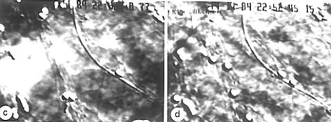
c - f. Subsequent photos: c. unprocessed picture, d. digital contrast enhancement, the single structures become apparent. The background fault remains.
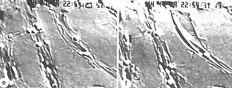
e. Subtraction of the background and resulting further contrast enhancement. f. the same, a few minutes later. The comparison of the particle distribution within the four pictures gives an impression of the plasma streaming velocity. The strands in e and f consist likewise mostly of ER. But clear indications exist that they are organized in parallel to the actin filament bundles along which small particle (here: sphaerosomes) move. (I. K. LICHTSCHEIDL and D. G. WEISS, 1988)