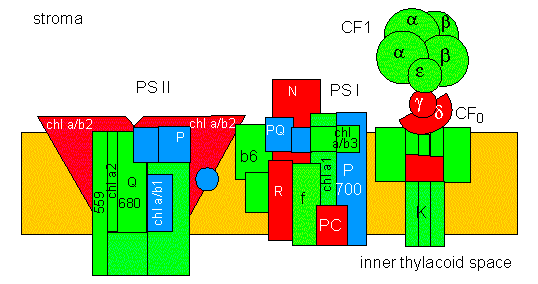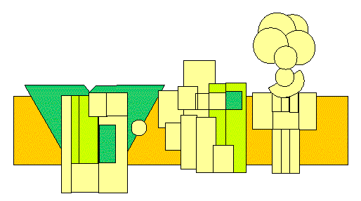
A model of the four complexes (PS II: photosystem II, cyt b6/f: part of the electron transport chain between PS II and PS I, PS I: photosystem I, CF1: ATP synthase, CF0: proton channel). The topology of some peptide subunits has been marked. Non-marked polypeptides are in literature characterized solely by their molecular weights: chl a/ b: chlorophyll a/b binding protein, chl a: chlorophyll a binding protein, 559: cytochrome 559, b6: cytochrome b6, f: cytochrome f, 700: reaction centre of PS I, 680: reaction centre of PS II, PQ: plastoquinone, PC: plastocyanin, FeS: iron-sulphur protein, R: Rieske-protein, N: NADP-binding protein. The subunits of CF0 have Greek letters as names, K: proton channel. Part A (at the top) of the picture: proteins encoded by the nucleus are blue, that of plastids have different colours. Part B (at the bottom) of the picture: chlorophyll-binding proteins are green (according to D. von WETTSTEIN and R. P. OLIVER, 1985).

© Peter v. Sengbusch - b-online@botanik.uni-hamburg.de