
Collenchyma Cell:
|
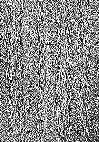
Electron microscopic picture of a cross-section through the cell wall of a collenchyma cell of Rumex conglomeratus, where the orientation of the microfibrils can be recognized in layers on top of each other (crossed position of the lamellas). The cellular axis corresponds to the vertical axis of the picture. The preparation has been shadowed. (S. C. CHAFE, 1970).