Lectures 16 and 17Muscle and motility
|
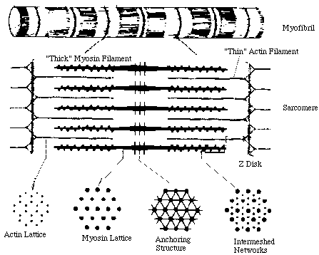
Muscle
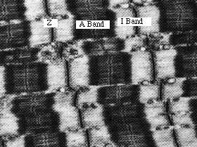
"Seen in longitudinal section (with an electron microscope), a myofilament shows several distict bands, each of which has been given a special letter. The lightest (least electron dense) band is known as the I band and consists mostly of actin. The wide, dark band, known as the A band, is composed primarily of myosin. In the center of the I band is an electron dense line, known as the Z-line. In the middle of the A band is another dense line known as the M line." (From Myofilament Structure link above).
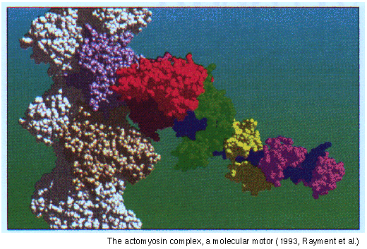
Myosin binding to actin (from Rayment et al.)
Myosin
Structure of myosin, and PDB files
2MYS.PDB, Structure of myosin head, and minor subunits.
1MMG.PDB, Structure of myosin head, with adenine nucleotide in situ, and conformational change.
The phylogeny of myosin
Actin
"At least six different types of actin are synthesized by vertebrate cells. The different genes which code them are highly conserved however, and they have similar properties. Globular, or G actin is stabilized by one tightly bound calcium ion. The protein is also associated with one molecule of ATP. The G actin polymerizes to form filamentous or F actin; this involves hydrolysis of the terminal phosphate of the ATP. Actin filaments consist of two strands of polymerized actin monomers, twisted into a helix with 13.5 molecules per turn." (From introduction to actin filaments reference below.)
Structure of actin, and PDB files
Actin-profilin complex (1hlu.pdb file)
Actin-DNA-ase complex (1atn.pdb file)
Muscle Links
Links to other muscle sites can be found on Washington State University Muscle page.
Cytoskeleton
Introduction to Cell Biology, including a nice page on microtubules, with extensive graphics.
A brief description of the role of the cytoskeleton in mitosis
Click here for a movie on the dynamic redistribution of the cytoskeleton during phagocytosis from Cells Alive.
"The cytoskeleton determines the shape of a cell, much like our bones help shape our bodies. In addition, the cytoskeleton makes the cell move, more like our muscles do. Like in muscle, this role is fulfilled in single cells in particular by the proteins actin and myosin. With the help of molecular genetics, a protein of the cytoskeleton called "coronin" that co-distributes with actin was labelled with green fluorescent protein, which is observable in cells like the living Dictyostelium discoideum shown above.
We have recorded the redistribution of coronin with a confocal microscope, which produces an image of a thin slice of the cell, like looking at only the beef within a hamburger. At regions which protrude from the surface of the cell, the so-called pseudopods, the labelled cytoskeletal protein accumulates transiently. This is also the case at a site where a yeast cell, marked with a red dye, is taken up by phagocytosis. After the particle is swallowed, the cytoskeletal protein returns to its original position beneath the cell surface." (From the Cells Alive page.)
Cilia and eukaryote flagella
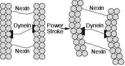
( Animation and Figs. from Molecular Machines)
A nice page on cilia and eukaryote flagella.
Chlamydomonas
The AXONEMAL DYNEIN family of proteins -- Summary
Kinesin
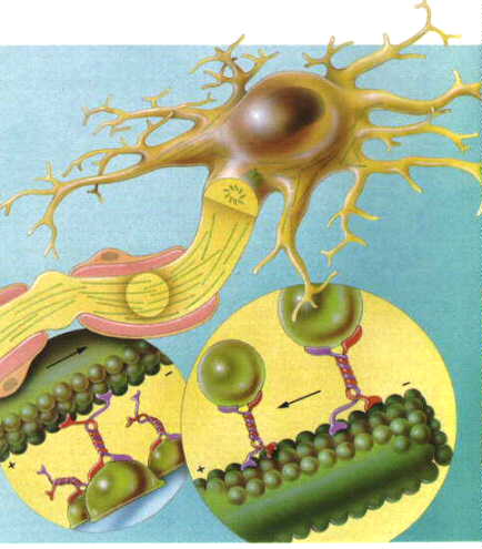
"3-D view of a neuron with its processes containing microtubules. At higher magnifications, the vesicles are seen attached to microtubule associated proteins (MAPs) and moving along the microtubule conveyer belt. The MAPs include kinesin and dynein which "walk" along the microtubules in opposite directions. Kinesin moves the vesicle along towards the plus end and dynein walks towards the minus end. In neurons, as the microtubules grow from the cell body through the processes, the plus end is more peripheral." (From the microtubules page above.)
Visit the kinesin Home Page, including information on the atomic structure of kinesin.
PDB files for some motility proteins
Bacterial motility and chemotaxis
All you want to know about bacterial motility and chemotaxis.
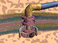
Flagella motor
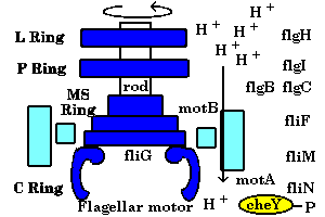
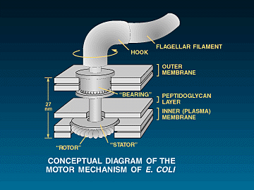
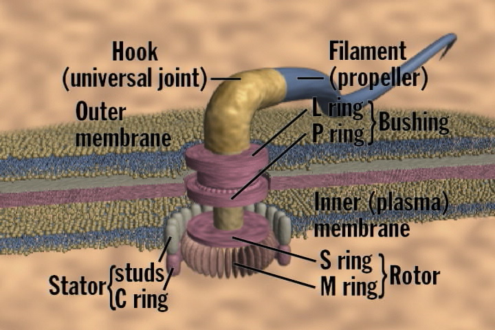
Animated motor
(Animation and Figs. from Molecular Machines)
Movie of E. coli swimming path (MPG)
Bacterial motility page from Cells Alive, with MOV movie
Bacterial sex

©Copyright 1996,
Antony Crofts, University of Illinois at Urbana-Champaign,
a-crofts@uiuc.edu