|
|
|
|
|
CELL WALLS |
|
|
|
|
Meristems: Sites of cell
division |
|
|
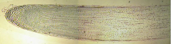
The root apical meristem (RAM) is
the simpler of the two apical meristems. The dividing cells
are just a little way behind the tip and they bud off cells
in two directions:
|
|
|
|
|
|
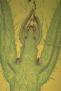
The shoot apical meristem is more complex. In addition to producing the stem it produces leaf and bud initials. The outer layers of cells tend to divide perpendicular to the surface and form the tunica whereas the inner cells of the corpus can divide in any direction. |
|
|
Apical meristems give rise to the primary meristems that form the tissues of the plant:
|
|
|
Other meristems arise later in plant growth, particularly in woody plants. The vascular cambium generates new xylem and phloem every year and the cork cambium produces the outer layers of bark. Mitosis |
|
|
The typical state for most cells is interphase. At the beginning of interphase (G1 phase) there is a single copy of the genetic material. If a cell is going to divide the DNA is replicated during the S phase. Then the cell is in the G2 phase with two copies of its DNA and ready to divide. |
|
|
In prophase the nuclear membrane breaks down, nucleoli disperse and chromosomes go from the extended configuration, typical of interphase to the condensed form. Each chromosome can be seen to be made up of two chromatids |
|
|
In metaphase the chromosomes line up on a"plate" about where the new cell wall will form; a spindle is organized from microtubules |
|
|
In anaphase the centromere of each chromosome splits and is drawn along the spindle fibers, taking a chromatid to opposite ends of the cell. |
|
|
In telophase the chromosomes cluster at each pole and the DNA becomes diffuse. The nuclear membrane and nucleoli reform; a cell plate begins to form between the new cells. |
|
|
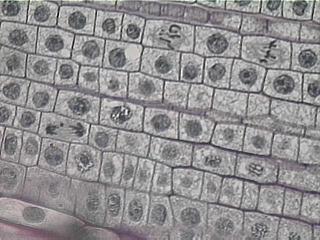
Stages of mitosis in an onion root tip. It is easiest to find nuclei in prophase, metaphase and telophase. You can see Virtual Mitosis online. |
|
|
Cell wall development |
|
|
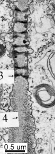
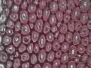
Part of the primary cell wall in a young strawberry fruit showing plasmodesmata (3). The relatively clear zone (4) in the middle of the wall is the middle lamella. The electron dense dots in the wall are cellulose microfibrils in section |
|
|
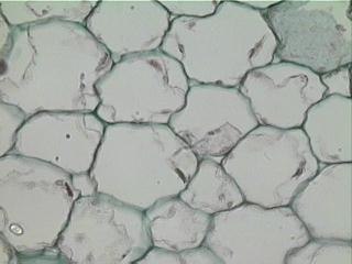
The epidermis and much of the ground tissue (pith and cortex), particularly in herbaceous plants, consists of parenchyma cells that have enlarged but undergone little differentiation. Parenchyma cells typically have thin, primary cell walls and most of their cell volume is occupied by vacuole. Leaf petioles and some stems are strengthened by collenchyma, parenchyma cells that develop thicker primary walls |
|
|
|
|
|
In other, more specialized cell types cell wall development continues with the deposition of the secondary wall. This has a higher proportion of cellulose and less pectin than the primary wall; secondary walls contain hemicelluloses (xyloglucan, xylan, mannan) in place of pectin and are often lignified. Thus when parenchyma cells have developed a lignified secondary wall they become sclerenchyma. Sclerenchyma cells can be globular sclereids as in the"stone cells" of pear fruits or elongated fibers which occur in many plant stems. Traditionally, fibers from flax, jute, hemp and sisal have been important for string, rope and woven fabrics, but these have been replaced to some extent by synthetic materials. |
|
|
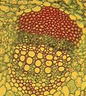
A transverse section of a vascular bundle in a Helianthus stem. The bundle is embedded in thin walled parenchyma cells. The thick red stained cell walls belong to fiber cells. The rows of large cells are vessel elements and between these and the fibers are phloem cells |
|
|
Copyright © Michael Knee, |
|