Bacterial Anatomy
Copyright 1998. Thomas M. Terry, University of Connecticut.
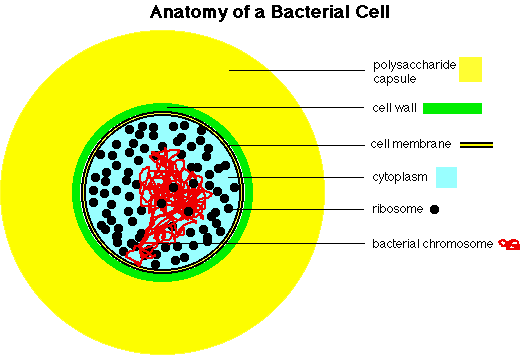
The figure below shows major anatomical structures found in most bacterial cells.

- Capsule: slimy layer, consisting of polysaccharide and water surrounding many cells. Also called slime coat, extracellular layer, etc. Difficult to stain, since it is mostly water.
- Cell Wall: rigid layer surrounding the bacterial cell. Made of peptidoglyan in bacteria, other materials in archaea. Porous to movement of small molecules.
- Cell Membrane: flexible, semi-permeable barrier with lipid center that controls diffusion in and out of cell.
- Cytoplasm: the fluid-filled space inside the cell. Contains hundreds of different enzymes, along with ribosomes, DNA, RNA, and a "pool" of millions of small molecules and ions.
- Ribosomes: particles made of protein and RNA, sites of protein assembly. Ribosomes may occupy 25% of the volume of a typical bacterial cell.
- Cell Chromosome: the DNA of a cell, normally a single circular molecule that is tightly supercoiled and packed inside the cell. Actively dividing cells may contain 2 or even 4 copies of this chromosome, replicated and ready for dividing up among future daughter cells.
