|
|
Quiz Me! |
Procaryote anatomy: cell envelope, motility, endospores |
|
|
|
Lecture Index | ||
|
|
Course Resources page |
Last revised: Wednesday, February 2, 2000
Ch. 3 (p. 51-69) in Prescott et al, Microbiology, 4th Ed. Note: These notes are provided as a guide to topics the instructor hopes to cover during lecture. Actual coverage will always differ somewhat from what is printed here. These notes are not a substitute for the actual lecture! Copyright 2000. Thomas M. Terry
Cell wall
- Rigid layer, preserves shape when rest of cell is digested.
- Made of peptidoglycan = polymer of peptides (typically 4 amino acids long, cross-linked to other chains) and glycans (made of alternating amino sugars)
- Sugars found in peptidoglycan
- N-acetylglucosamine (NAG).
- N-acetylmuramic acid (NAM).
- NAG-NAM sugars are linked by ß-1,4 linkage). See Fig. 3.20-3.29 in texts. Note: glycan chain is similar to chitin (= lobster shell), a polymer of NAG.
chemical structure of peptidoglycan (symbols: G = NAG, M = NAM)
Unusual properties of PG:
- contains D-amino acids (proteins usually made only of L-amino acids), such as D-alanine, D-glutamic acid; also unusual amino acid diaminopimelic acid;
- synthesized not on ribosomes, but by series of enzymes
Mechanism of synthesis of Peptidoglycan
- (NOTE: analogous to cutting open tire, adding more rubber, sealing up, without losing pressure!)
- enzyme needed to cut PG = autolysin
- two Carrier molecules: uridine diphosphate (related precursor of RNA synthesis also) and bactoprenol (55-C atom alcohol)
- synthesis begins inside cell with construction of NAG-NAM-peptide unit.
- carry across membrane, insert into growing wall.
- PENICILLIN: blocks cross peptide linkage. Wall loses all rigidity (only affects growing cells).
- View effects of penicillin on bacteria
Structural Basis for the Gram Stain
- Christian Gram recognized two different types of bacteria based on reaction to certain staining procedure.

- View cartoon diagram of gram+ and gram- cell wall
- With EM, could see that Gram+ cells have thicker walls.
- Gram+ cells have thick peptidoglycan layer, also have teichoic acids = polymers of polyalcohols (ribitol or glycerol) + phosphate; also attached D-ala or sugars. Function not clear: highly - charged, probably project out through PG layer.
- Gram- cells have much thinner peptidoglycan, but also have outer membrane (OM) made of lipid, protein, and LPS (lipopolysaccharide). OM is porous because of porin proteins that create pores, allow small molecules to pass freely.
- Periplasmic space = space between Inner membrane and OM. May occupy as much as 30% of cell volume. Not just an entryway: contains many specialized proteins, often involved in nutrient acquisition. Ex: phosphatases, enzymes that cleave organic molecules containing phosphate, make it available for cell nutrition.
- Can "shock" periplasmic proteins out by sudden immersion in ice cold water.
Osmotic effects
- Water moves freely across membranes (= osmosis) to try to equalize concentration. Interior of cell = ~ 0.9% salt.
- If surrounding fluid = 0.9% salt, Isotonic. Equal water flow in & out
- If surrounding fluid < 0.9% salt, Hypotonic. Water flows in, presses against wall. No problem if wall is intact (can withstand 20 atmospheres of pressure, vs 2 for car tire)
- If surrounding fluid > 0.9% salt, Hypertonic. Water flows out; cell shrinks --> plasmolysis. Stops metabolism.
Effects of lysozyme and pencillin
Challenge Question
- Growing cell must break peptidoglyan at certain spots, insert new material, seal back together to grow new wall
- Cell wall contains bacterial enzymes called autolysins to make these breaks, but carefully controlled. Mechanism not well understood.
- Lysozyme = enzyme that attacks PG, breaks glycan chain. Found in animal secretions, e.g. tears, saliva, mucous membranes. More active against Gram+ than Gram- cells.
- Penicillin = fungal antibiotic, blocks formation of new peptide links in growing cells walls (= transpeptidation). Note particular advantage of penicillin: no harmful effects on human cells, since PG is unique to bacteria. Penicillin G (the natural isolate) is less effective against many gram-negative cells, since it has difficulty penetrating the outer membrane.
Spheroplasts, Protoplasts L-forms, and Mycoplasmas
- Lysozyme and penicillin can lead to cells without walls. Cells lack rigid shape, assume spherical shape = spheroplast (if gram-negative cell) or protoplast (if gram-positive parent). Still able to live if isotonic, but will lyse if put in hypotonic environment.
- L-forms = stable spheroplasts, grow indefinitely w/o wall, but derived from parents with normal walls.
- Mycoplasmas = group of bacteria that evolved to lack wall. Typically found in environments with high osmotic strength (e.g. sewage, vagina. Names like M. orales, M. genitalium, M. pneumoniae.
Archaea: different cell walls
- Cell walls in certain bacteria made of other materials, not peptidoglycan. Can be protein, glycoprotein, or polysaccharide. Some archaea have pseudopeptidoglycan, similar architecture, but N-acetyltalosaminuronic acid instead of N-acetylmuramic. All archaeal walls are resistant to lysozyme.
- These groups collectively called Archaea (because habitats resemble those common of primitive earth); appear to be as different from other bacteria (Eubacteria) as either are from Eukaryotes!
What are the Archaea?
- When the distinction between prokaryotes and eukaryotes was made as a result of extensive electron micrographic studies of cell types (~ 1940's and 1950's), it was at first thought that all bacteria (prokaryotes) were fundamentally more similar to each other than to eukaryotes.
- However, certain groups of prokaryotes turned out do differ profoundly from the rest. These organisms fell into four broad groups:
- Methanogens; anaerobic methane producers
- Extreme Halophiles; salt-dependent organisms
- Hyperthermophiles; heat-dependent organisms
- Thermoplasma; a group of heat and acid-dependent organisms lacking cell walls
- Because many of these habitats resembled conditions of the early (archaic) earth, such as hot, acidic, or salty, these organisms were collectively called Archaea. The remaining bacteria were collectively called the Bacteria (sometimes referred to as Eubacteria, or "true bacteria").
- We now recognize that the Archea are taxonomically as different from Bacteria as either are from eukaryotes, which collectively are called Eukarya.
- These three groups of organisms are called the 3 Domains of Life:
- Archaea
- Bacteria
- Eukarya
Structural Differences between Archaea and Bacteria
- Cell Wall Architecture
- Bacterial walls are made of peptidoglycan, a polymer of N-acetyl glucosamine and N-acetyl muramic acid (glycan chain) with short peptides containing both D- and L-amino acids.
- Archaeal walls differ widely, and are made from different materials. Walls of methanogens often contain pseudopeptidoglycan, similar to peptidoglycan, but with slightly different sugars and archictecture. Other archaea use polysaccharides, proteins, or glycoproteins as wall materials.
- Many eukaryotes have cell walls. These are built of a variety of materials, but never peptidoglycan.
- Fatty Acid Linkages
- In Bacteria and Eukaryotes, membrane fatty acids are linked to glycerol by ester bonds.
- In Archaea, membranes are built from different types of lipids, polymers of the highly unsaturated molecule isoprene. These lipids are linked by ether bonds, not ester bonds.
- Structure of RNA Polymerase
- RNA polymerase is a crucial enzyme required for the synthesis of new RNA molecules. In bacteria, there is a single type of this enzyme, and it is built of four subunits.
- In eukaryotes, there are three different enzymes, and they each possess 8-12 subunits.
- Archaea have intermediate properties; they have only a single enzyme, like bacteria, but it is made of 8-12 subunits, like eukaryotes.
- Initiation Codon
- Proteins are synthesized on ribosomes, with the precise sequence of amino acids dictated by the genetic code.
- Ribosomes recognize a unique codon, called the initiation codon (AUG), as the correct location to begin synthesizing a protein.
- In eukaryotes and archaea, the AUG codon always specifies the amino acid methionine
- In Bacteria, the AUG codon specifies N-formylmethionine, a modified form of methionine.
Table summarizing differences between Bacteria and Archaea
Property Bacteria Archaea Eukarya Cell wall Made of peptidoglyan Made of various materials, not peptidoglyan (If present) cellulose, others Lipids Fatty acids present, linked by ester bonds Isoprenes present, linked by ether bonds Fatty acids present, linked by ester bonds RNA polymerase enzyme Single small enzyme; 4 subunits Single large enzyme; many subunits Three large enzymes; many subunits Protein synthesis 1st amino acid = formylmethionine 1st amino acid = methionine 1st amino acid = methionine
Outside the envelope
- glycocalyx (also called slime layer, capsule). Not found in all bacteria.
- varies in thickness, rigidity.
- important in adhesion, ability to avoid phagocytosis
- some suggestion that many bacteria lose layer when cultured in laboratory.
- May be much more prominent in nature than thought.
- Bacterial adhesion promotes formation of biofilms, masses of bacteria encased in large aggregates of extracellular matrix. Biofilms are not well-understood, but incredibly important. Most bacteria may live largely in biofilms rather than as free organisms (the dispersal stage). Biofilms are harder to get rid of, more resistant to antibiotics.
- fimbriae & pili
- short, rigid protein rods, similar in size to flagella, but not involved in motility.
- function in adhesion, formation of pellicles at liquid surfaces. Function not entirely clear. Pili sometimes involved in pathogenic adhesion (e.g. gonnorhaea)
- View electron micrograph of E. coli with frimbriae
- Flagella
- curved filament made of flagellin protein: travels through hollow tube, assembles at external end.
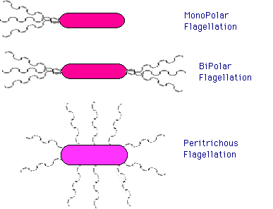
- can be arranged in two ways:

- polar flagellation: flagella attached at one (monopolar) or both (bipolar) ends. Ex: Pseudomonas aeruginosa
- peritrichous flagellation: flagella attached at many sites around cell periphery. Ex: E. coli
- attached to cell via basal region
- flagellar rotor can rotate in either direction: CW or CCW. Signals from cell control direction of rotation. See Motility below for application
- flagellar rotor is the only circular rotor found in nature, aside from human artifact
Motility
- Flagellar Motility & Chemotaxis
- View Bacterial motility from "Cells Alive"
- Flagella can rotate clockwise (CW) -- in peritrichous cells, flagella then become limp, cell TUMBLES or TWIDDLES -- or countercloskwise (CCW) -- flagellar bundle then becomes rigid, cell RUNS. Rotor is always spinning one direction or other.
- Energy for rotation comes from Proton gradient.
- flagellated bacteria move through gradients, TOWARD ATTRACTANTS, AWAY from REPELLANTS.
- How? detect temporal gradient. If moving towards attractant, suppress tumbles. If moving away, increase frequ. of tumbles.
- complex mechanism in cell membrane: (1) protein receptors bind chemical; (2) membrane proteins (Methyl-accepting chemotaxis proteins) transmit signal.
- Other forms of motility
- some bacteria are motile w/o flagella. GLIDING MOTILITY. move slowly across surfaces, involves sulfur-containing lipids.
Structural adaptations to inhospitable environments
- Many bacteria can survive for extended periods in absence of food, water, conditions for growth.
- One adaptation: cells entering poor environments shut down normal metabolic pathways, make many new proteins to protect cell. Can survive extended periods of dehydration.
- Some bacteria produce dormant structures: spores, cysts. Allow prolonged survival, dispersal.
- A few genera of bacteria produce highly specialized survival structures = Endospores.
- Major source of difficulty with sterilization. Some resist boiling up to several hours.
- Spore is inert storage form. Found in certain species, most abundantly Bacillus and Clostridia. Formed when cells age, run out of nutrients.
- Very refractile, difficult to stain. Coated with thick layers, reduced water content. All cell materials present inside coats, but metabolically inactive.
- Spore coat conatins DIPICOLINIC ACID (DPA) and high Ca. Responsible for part of heat-resistance
- Germination can happen in minutes: spore loses refractility, swells visibly, vegetative cell pushes out of coat.
Take a Self-Quiz on this material
Return to Lecture Index
Return to MCB 229 Course Resources page