 Go to the
top page
Go to the
top page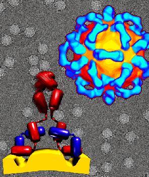
FIGURE 1: (TOP) Cryoelectron micrograph (gray) and computer enhanced image (in color) of spherical human rhinovirus 14 particles (yellow &orange) saturated with a neutralizing monoclonal antibody (blue) (mAb17 IgG2a, directed against the NIM-IA site). The particle is assembled from 12 pentameric subunits. The Fc portion of the IgG is not visible, apparently because it is too mobile to produce reinforcement in the enhancement stage. This is the first direct evidence in support of the pentamer bridging hypothesis proposed by Mosser et al. in 1989 (A morph animation illustrates corresponding density in a previous, inconclusive 3D cryoelectron microscopy dataset). Pentamer bridging by an antibody molecule is illustrated by an animated model.
(BOTTOM) Each bivalent IgG molecule (red, heavy chains, purple,light chains) bridges two canyons on adjacent pentamers (yellow) of the virus shell. (The hypothesized correspondance of the IgG model to the 3D cryoelectron microscopy image reconstruction is shown by a morph animation.) Binding of IgG molecules inhibits attachment by preventing insertion of cellular receptor into the canyon. Mobility of the Fc region is indicated by blurring in the region farthest from the virus surface.
Photo courtesy Prof. Tom Smith (tom@bragg.bio.purdue.edu) Purdue University.
Go to the
top page
© 1994 Tom Smith (tom@bragg.bio.purdue.edu). Purdue University / tom@bragg.bio.purdue.edu