
Digestive System | Plans and Locations | Stages in the Digestive Process
Components of the Digestive System | Regulation of Appetite | Nutrition | Links
Single-celled organisms can directly take in nutrients from their outside environment. Multicellular animals, with most of their cells removed from contact directly with the outside environment, have developed specialized structures for obtaining and breaking down their food. Animals depend on two processes: feeding and digestion.
Animals are heterotrophs, they must absorb nutrients or ingest food sources. Ingestive eaters, the majority of animals, use a mouth to ingest food. Absorptive feeders, such as tapeworms, live in a digestive system of another animal and absorb nutrients from that animal directly through their body wall. Filter feeders, such as oysters and mussels, collect small organisms and particles from the surrounding water. Substrate feeders, such as earthworms and termites, eat the material (dirt or wood) they burrow through. Fluid feeders, such as aphids, pierce the body of a plant or animal and withdraw fluids.
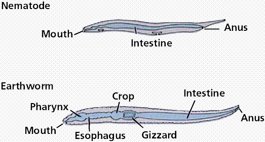
The above images are modified from http://www.whfreeman.com/life/update/.
The digestive system uses mechanical and chemical methods to break food down into nutrient molecules that can be absorbed into the blood.
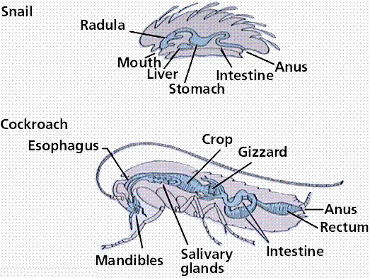
There are two types of plans and two locations of digestion. Sac-like plans are found in many invertebrates, who have a single opening for food intake and the discharge of wastes. Vertebrates use the more efficient tube-within-a-tube plan with food entering through one opening (the mouth) and wastes leaving through another (the anus).
Intracellular digestion: food is taken into cells by phagocytosis with digestive enzymes being secreted into the phagocytic vesicles; occurs in sponges, coelenterates and most protozoans.
Extracellular digestion: digestion occurs in the lumen (opening) of the digestive system, with the nutrient molecules being transferred to the blood or body fluid; occurs in chordates, annelids, and crustaceans.
Three phases occur during what we loosely refer to as "digestion". Digestion proper, which is the mechanical and chemical breakdown of food into particles/molecules small enough to pass into the blood stream. Absorption into the blood stream. Assimilation, the passage of the food molecules into body cells.
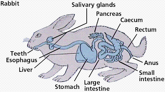
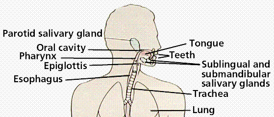
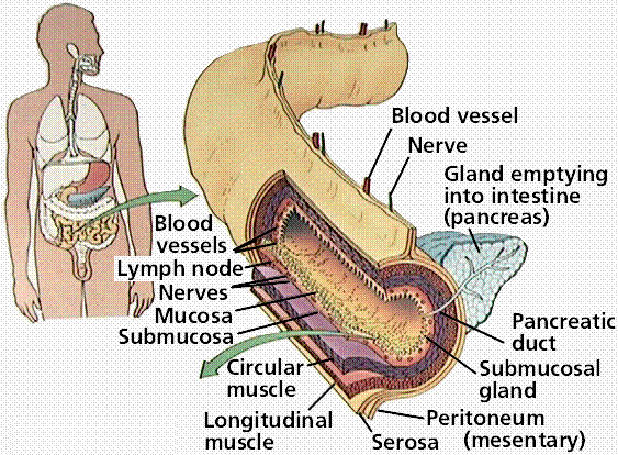
The human digestive system is a coiled, muscular tube (6-9 meters long when fully extended) extending from the mouth to the anus. Several specialized compartments occur along this length: mouth, pharynx, esophagus, stomach, small intestine, large intestine, and anus. Accessory digestive organs are connected to the main system by a series of ducts: salivary glands, parts of the pancreas, and the liver and gall bladder (bilary system).
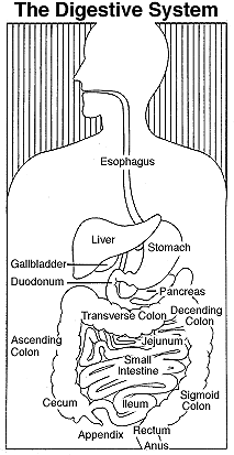
The above image is from http://www.niddk.nih.gov/digesyst/digesyst.html.
The above images are modified from http://www.whfreeman.com/life/update/.
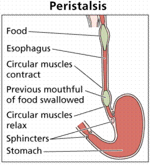
Mechanical breakdown begins in the mouth by chewing (teeth) and actions of the tongue. Chemical breakdown of starch by production of salivary amylase from the salivary glands. This mixture of food and saliva is then pushed into the pharynx and esophagus. The esophagus is a muscular tube whose muscular contractions (peristalsis) propel food to the stomach.
In the mouth, teeth, jaws and the tongue begin the mechanical breakdown of food into smaller particles. Most vertebrates, except birds (who have lost their teeth to a hardened bill), have teeth for tearing, grinding and chewing food. The tongue manipulates food during chewing and swallowing; mammals have tastebuds clustered on their tongues.
Salivary glands secrete salivary amylase, an enzyme that begins the breakdown of starch into glucose. Mucus moistens food and lubricates the esophagus. Bicarbonate ions in saliva neutralize the acids in foods.
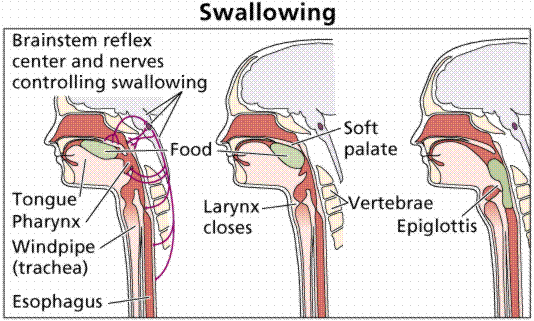
Swallowing moves food from the mouth through the pharynx into the esophagus and then to the stomach.
The above image is modified from http://www.whfreeman.com/life/update/.
The above image is modified from http://www.whfreeman.com/life/update/.
During a meal, the stomach gradually fills to a capacity of 1 liter, from an empty capacity of 50-100 milliliters. At a price of discomfort, the stomach can distend to hold 2 liters or more.
Epithelial cells line inner surface of the stomach, and secrete about 2 liters of gastric juices per day. Gastric juice contains hydrochloric acid, pepsinogen, and mucus; ingredients important in digestion. Secretions are controlled by nervous (smells, thoughts, and caffeine) and endocrine signals. The stomach secretes hydrochloric acid and pepsin. Hydrochloric acid (HCl) lowers pH of the stomach so pepsin is activated. Pepsin is an enzyme that controls the hydrolysis of proteins into peptides. The stomach also mechanically churns the food. Chyme, the mix of acid and food in the stomach, leaves the stomach and enters the small intestine.
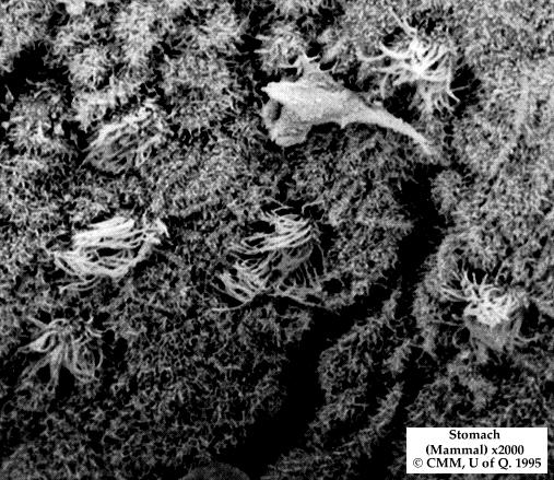
This image is from http://130.102.208.100/FMRes/FMPro?-db=images.fp3&key=32819&-img, used by permission of Nanoworld. Scanning electron micrograph of the stomach lining of a mammal, X2000.
Hydrochloric acid does not directly function in digestion: it kills microorganisms, lowers the stomach pH to between 1.5 and 2.5; and activates pepsinogen. Pepsinogen is an enzyme that starts protein digestion. Pepsinogen is produced in cells that line the gastric pits. It is activated by cleaving off a portion of the molecule, producing the enzyme pepsin that splits off fragments of peptides from a protein molecule during digestion in the stomach.
Carbohydrate digestion, begun by salivary amylase in the mouth, continues in the bolus as it passes to the stomach. The bolus is broken down into acid chyme in the lower third of the stomach, allowing the stomach's acidity to inhibit further carbohydrate breakdown. Protein digestion by pepsin begins.
Alcohol and aspirin are absorbed through the stomach lining into the blood.
Epithelial cells secrete mucus that forms a protective barrier between the cells and the stomach acids. Pepsin is inactivated when it comes into contact with the mucus. Bicarbonate ions reduce acidity near the cells lining the stomach. Tight junctions link the epithelial stomach-lining cells together, further reducing or preventing stomach acids from passing.
Peptic ulcers result when these protective mechanisms fail. Bleeding ulcers result when tissue damage is so severe that bleeding occurs into the stomach. Perforated ulcers are life-threatening situations where a hole has formed in the stomach wall. At least 90% of all peptic ulcers are caused by Helicobacter pylori. Other factors, including stress and aspirin, can also produce ulcers.
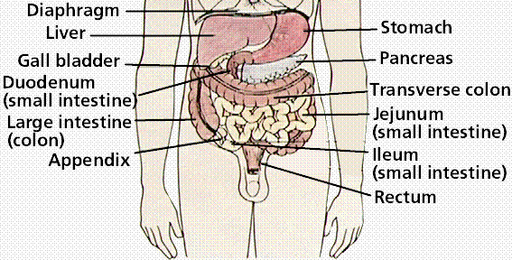
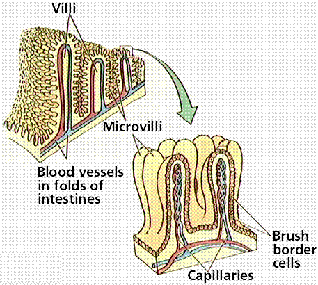
The small intestine is where final digestion and absorption occur. The small intestine is a coiled tube over 3 meters long. Coils and folding plus villi give this 3m tube the surface area of a 500-600m long tube. Final digestion of proteins and carbohydrates must occur, and fats have not yet been digested. Villi have cells that produce intestinal enzymes which complete the digestion of peptides and sugars. The absorption process also occurs in the small intestine. Food has been broken down into particles small enough to pass into the small intestine. Sugars and amino acids go into the bloodstream via capillaries in each villus. Glycerol and fatty acids go into the lymphatic system. Absorption is an active transport, requiring cellular energy.
The above images are modified from http://www.whfreeman.com/life/update/.
Food is mixed in the lower part of the stomach by peristaltic waves that also propel the acid-chyme mixture against the pyloric sphincter. Increased contractions of the stomach push the food through the sphincter and into the small intestine as the stomach eempties over a 1 to 2 hour period. High fat diets significantly increase this time period.
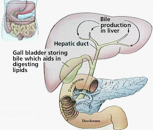
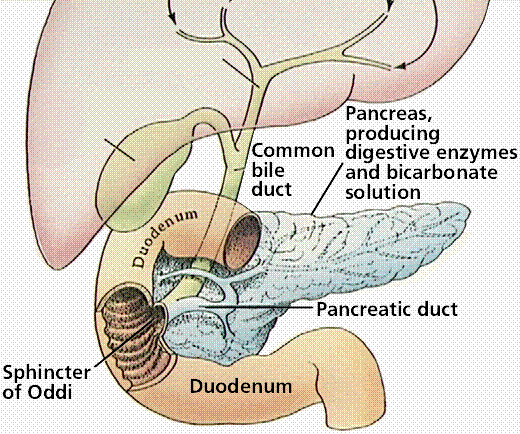
The small intestine is the major site for digestion and absorption of nutrients. The small intestine is up to 6 meters long and is 2-3 centimeters wide. The upper part, the duodenum, is the most active in digestion. Secretions from the liver and pancreas are used for digestion in the duodenum. Epithelial cells of the duodenum secrete a watery mucus. The pancreas secretes digestive enzymes and stomach acid-neutralizing bicarbonate. The liver produces bile, which is stored in the gall bladder before entering the bile duct into the duodenum.
Digestion of carbohydrates, proteins, and fats continues in the small intestine. Starch and glycogen are broken down into maltose. Proteases (enzymes secreted from the pancreas) continue the breakdown of protein into small peptide fragments and some amino acids.
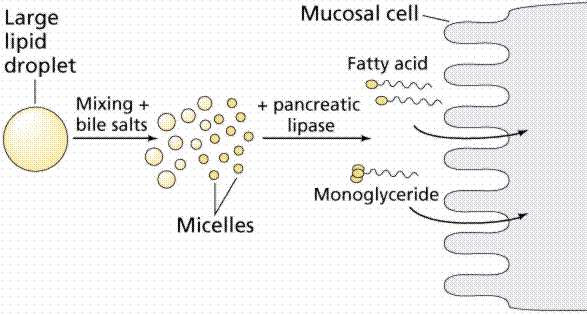
Bile emulsifies fats, facilitating their breakdown into progressively smaller fat globules until they can be acted upon by lipases. Bile contains cholesterol, phospholipids, bilirubin, and a mix of salts. Fats are completely digested in the small intestine, unlike carbohydrates and proteins.
Most absorption occurs in the duodenum and jejeunum (second third of the small intestine). The inner surface of the intestine has circular folds that more than triple the surface area for absorption. Villi covered with epithelial cells increase the surface area by another factor of 10. The epithelial cells are lined with microvilli that further increase the surface area; a 6 meter long tube has a surface area of 300 square meters.
Each villus has a surface that is adjacent to the inside of the small intestinal opening covered in microvilli that form on top of an epithelial cell known as a brush border. Each villus has a capillary network supplied by a small arteriole. Absorbed substances pass through the brush border into the capillary, usually by passive transport.
Maltose, sucrose, and lactose are the main carbohydrates present in the small intestine; they are absorbed by the microvilli. Starch is broken down into two-glucose units (maltose) elsewhere. Enzymes in the cells convert these disaccharides into monosaccharides that then leave the cell and enter the capillary. Lactose intolerance results from the genetic lack of the enzyme lactase produced by the intestinal cells.
Peptide fragments and amino acids cross the epithelial cell membranes by active transport. Inside the cell they are broken into amino acids that then enter the capillary. Gluten enteropathy is the inability to absorb gluten, a protein found in wheat.
Digested fats are not very soluble. Bile salts surround fats to form micelles that can pass into the epithelial cells. The bile salts return to the lumen to repeat the process. Fat digestion is usually completed by the time the food reaches the ileum (lower third) of the small intestine. Bile salts are in turn absorbed in the ileum and are recycled by the liver and gall bladder. Fats pass from the epithelial cells to the small lymph vessel that also runs through the villus.
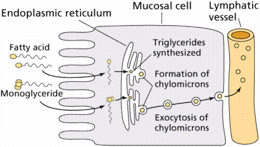
The above image is modified from http://www.whfreeman.com/life/update/.
The liver sends bile to the small intestine. Bile contains bile salts, which emulsify fats, making them susceptible to enzymatic breakdown. In addition to digestive functions, the liver functions in other systems: 1) detoxification of blood; 2) synthesis of blood proteins; 3) destruction of old erythrocytes and conversion of hemoglobin into a component of bile; 4) production of bile; 5) storage of glucose as glycogen; and 6) production of urea from amino groups and ammonia.
The above images are modified from http://www.whfreeman.com/life/update/.
Glycogen (chains of glucose molecules) serves as a reservoir for glucose. Low glucose levels in the blood cause release of hormones to stimulate breakdown of glycogen into glucose. When no glucose or glycogen is available, amino acids are converted into glucose in the liver. The process of deamination removes the amino groups from amino acids. Urea is formed and passed through the blood to the kidney for export from the body.
Jaundice occurs when the characteristic yellow tint to the skin is caused by excess hemoglobin breakdown products in the blood, a sign that the liver is not properly functioning. Jaundice may occur when liver function has been impaired by obstruction of the bile duct and by damage caused by hepatitis. Hepatitis A, B, and C can all cause liver damage. Cirrhosis of the liver commonly occurs in alcoholics, who place the liver in a stress situation due to the amount of alcohol to be broken down.
The pancreas sends pancreatic juice, which neutralizes the chyme, to the small intestive through the pancreatic duct.
The large intestine is made up by the colon, cecum, appendix, and rectum. Material in the large intestine is mostly indigestible residue and liquid. Movements are due to involuntary contractions that shuffle contents back and forth and propulsive contractions that move material through the large intestine.
Secretions in the large intestine are an alkaline mucus that protects epithelial tissues and neutralizes acids produced by bacterial metabolism. Water, salts, and vitamins are absorbed, the remaining contents in the lumen form feces (mostly cellulose, bacteria, bilirubin). Bacteria in the large intestine, such as E. coli, produce vitamins (including vitamin K) that are absorbed.
The hypothalamus in the brain has two centers controlling hunger. One is the appetite center, the other the satiety center.
Gastrin, secretin, and cholecystokinin are hormones that regulate stages of digestion. Protein in the stomach stimulates secretion of gastrin, which causes increased stomach acid secretion and mobility of the digestive tract to move food. Food passing into the duodenum causes the production of secretin, which in turn promotes release of alkaline secretions from the pancreas, stops further passage of food into the intestine until the acid is neutralized. Cholecystokinin (CCK) is released from intestinal epithelium in response to fats, and causes the release of bile from the gall bladder and lipase (a fat digesting enzyme) from the pancreas.
Glucose levels in the blood remain fairly stable. The liver absorbs glucose from the blood and stores it as the polysaccharide glycogen. Blood glucose levels are maintained between meals by releasing glucose from glycogen back into the blood.
Nutrition deals with the composition of food, its energy content, and slowly (or not at all) synthesized organic molecules. Chemotrophs are organisms (mostly bacteria) deriving their energy from inorganic chemical reactions. Phototrophs convert sunlight energy into sugar or other organic molecules. Heterotrophs eat to obtain energy from the breakdown of organic molecules in their food.
Macronutrients are foods required on a large scale each day. These include carbohydrates, lipids, and amino acids. Water is essential, correct water balance is a must for proper functioning of the body.
About 60% of the diet should be carbohydrates, such as are in milk, meat, vegetables, grains and grain products. The diet should contain at least 100 grams of carbohydrate every day.
Proteins are polymers composed of amino acids. Proteins are found in meat, milk, poultry, fish, cereal grains and beans. They are needed for cellular growth and repair. Twenty amino acids are found in proteins, of which humans can make eleven. The remaining nine are the essential amino acids which must be supplied in the diet. Normally proteins are not used for energy, however during starvation muscle proteins are broken down for energy. Excess protein can be used for energy or converted to fats.
Lipids and fats generate the greatest energy yield, so many plants and animals store energy as fats. Lipids and fats are present in oils, meats, butter, and plants (such as avocado and peanuts). Some fatty acids, such as linoleic acid, are essential and must be included in the diet. When present in the intestine, lipids promote the uptake of vitamins A, D, E, and K.
Vitamins are organic molecules required for metabolic reactions. They usually cannot be made by the body and are needed in trace amounts. Vitamins may act as enzyme cofactors or coenzymes. Some vitamins are soluble in fats, some in water.
Minerals are trace elements required for normal metabolism, as components of cells and tissues, and for nerve conduction and muscle contraction. They can only be obtained from the diet. Iron (for hemoglobin), iodine (for thyroxin), calcium (for bones), and sodium (nerve message transmission) are examples of minerals.
There is a quantitative relationship between nutrients and health. Imbalances can cause disease. Many studies have concluded nutrition is a major factor in cardiovascular disease, hypertension, and cancer.
Back to Table of Contents | THE NERVOUS SYSTEM
Email: mj.farabee@emcmail.maricopa.edu![]()
Last modified: 2000/01/05:08:59:28
The URL of this page is: gened.emc.maricopa.edu/bio/BIO181/BIOBK/BioBookDIGEST.html