
Monera: the Prokaryotic Kingdom | Bacterial Structure | Bacterial Reproduction
Classification of Bacteria | The Archea | The Fossil Record | Links
The taxonomic Kingdom Monera consists of the bacteria (meaning the true bacteria and cyanobacteria, or photosynthetic bacteria). Organisms in this group lack membrane-bound organelles associated with higher forms of life. Such organisms are known as prokaryotes. Bacteria (technically the Eubacteria) and blue-green bacteria (the blue-green algae when I was a student), or cyanobacteria are the major forms of life in this kingdom. The most primitive group, the archaebacteria, are today restricted to marginal habitats such as hot springs or areas of low oxygen concentration. Their small size, ability to rapidly reproduce (E. coli can reproduce by binary fission every 15 minutes), and diverse habitats/modes of existence make monerans the most abundant and diversified kingdom on Earth. Bacteria occur in almost every environment on Earth, from the bottom of the ocean floor, deep inside solid rock, to the cooling jackets of nuclear reactors. Possible bacteria-like structures have even been recovered from 3 billion year old Martian meteorites. If these turn out to be fossils, then the bacterial form of life would have existed simultaneously on both Earth and Mars. However, the cellular nature of those structures has not been conclusively established.
The above left image is cropped from gopher://wiscinfo.wisc.edu:2070/I9/.image/.bot/.130/Cyanobacteria/Oscillatoria_130. The above image right is cropped from gopher://wiscinfo.wisc.edu:2070/I9/.image/.bot/.130/Cyanobacteria/Nostoc_130.
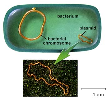
Bacteria lack a nuclear membrane and membrane-bound organelles. Biochemical processes that normally occur in a choloroplast or mitochondrion of eukaryotes will take place in the cytoplasm of prokaryotes. Bacterial DNA is circular and arrayed in a region of the cell known as the nucleoid. Scattered within bacterial cytoplasm are numerous small loops of DNA known as plasmids. Bacterial genes are organized in by gene systems known as operons.
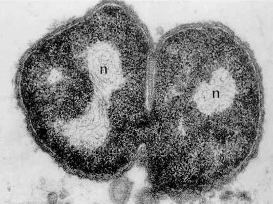
This image is from: http://129.109.136.65/microbook/ch002.htm Note the nucleoid region (n) where DNA is located as well as the electron dense areas of the cytoplasm (dark areas) on these two cells of Neisseria gonorrhoeae.
Structure of a "typical" bacterium. Image from W.H. Freeman and Sinauer Associates, used by permission.
Plasmids, small DNA fragments, are known from almost all bacterial cells. Plasmids carry between 2 and 30 genes. Some seem to have the ability to move in and out of the bacterial chromosome.
The above is from http://www.biosci.uga.edu/almanac/bio_103/notes/may_30.html.
The operon model of prokaryotic gene regulation was proposed by Fancois Jacob and Jacques Monod. Groups of genes coding for related proteins are arranged in units known as operons. An operon consists of an operator, promoter, regulator, and structural genes. The regulator gene codes for a repressor protein that binds to the operator, obstructing the promoter (thus, transcription) of the structural genes. The regulator does not have to be adjacent to other genes in the operon. If the repressor protein is removed, transcription may occur.
Structure of a typical operon. Image from W.H. Freeman and Sinauer Associates, used by permission.
Operons are either inducible or repressible according to the control mechanism. Seventy-five different operons controlling 250 structural genes have been identified for E. coli.
Bacteria have flagella with a different microtubule structure than the flagella of eukaryotes. Cell walls of bacteria contain peptidoglycan instead of the cellulose found in cell walls of plants and some algae. Ribosomes are the structures in cells where proteins are assembled. Bacterial ribosomes have different sized ribosomal subunits than do eukaryotes.
This image is from: http://www.slic2.wsu.edu:82/hurlbert/micro101/pages/Chap3.html
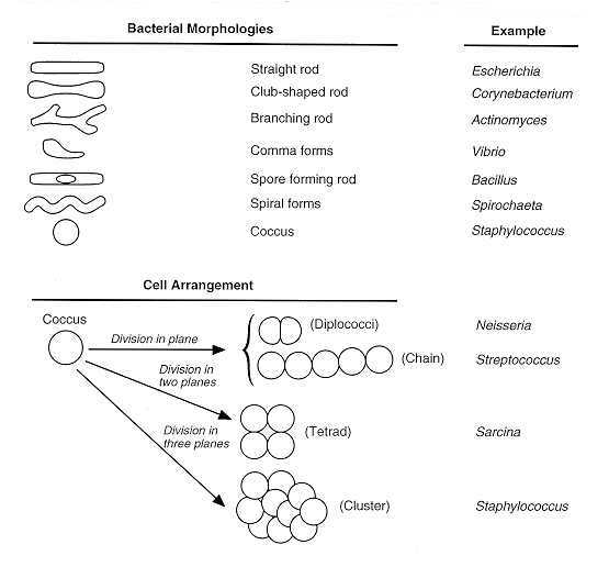
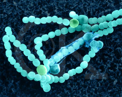
Bacteria typically have one of three shapes: rods (bacilli), spheres (cocci) or spiral (spirilla). Unicellular, they often stick together forming clumps or filaments.
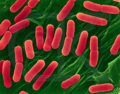
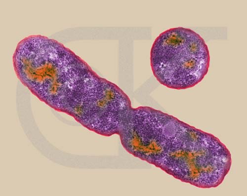
Rod-Shaped Bacterium, hemorrhagic E. coli, strain 0157:H7 (division) (SEM x22,810). This image is copyright Dennis Kunkel http://www.pbrc.hawaii.edu/~kunkel/gallery, used with permission.
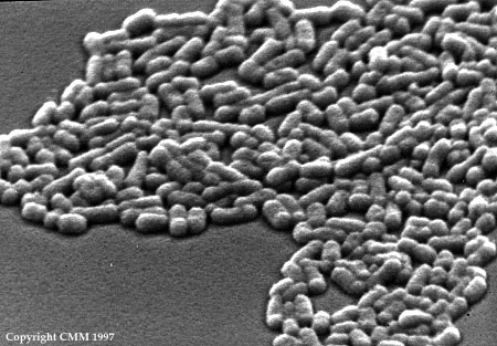
The top image above is from: http://www.uq.oz.au/nanoworld. The lower image above is from: http://www.slic2.wsu.edu:82/hurlbert/micro101/pages/Chap2.html#two_bact_groups Both illustrate the rod-shaped bacterium E. coli.
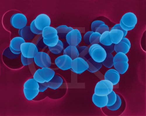
Coccoid-shaped Bacterium(causes skin infections), Enterococcus faecium (SEM x33,370). This image is copyright Dennis Kunkel http://www.pbrc.hawaii.edu/~kunkel/gallery, used with permission.
The image above is from http://www.bact.wisc.edu/Bact303/MajorGroupsOfProkaryotes. Left, a cross-section of a cell illustrating the location of a flagella inside the cell; Center, Borrelia burgdorferi, the organism that causes Lyme disease; and Right, Treponema pallidum, the spirochete that causes the venereal disease syphilis.
This image is from: http://129.109.136.65/microbook/ch002.htm
Some bacteria are photosynthetic autotrophs, others are heterotrophs.
Prokaryotes are much simpler in their organization than are eukaryotes. There are a great many more organelles in eukaryotes, also more chromosomes. The usual method of prokaryote cell division is binary fission. The prokaryotic chromosome is a single DNA molecule that first replicates, then attaches each copy to a different part of the cell membrane. When the cell begins to pull apart, the replicate and original chromosomes are separated. Following cell splitting (cytokinesis), there are then two cells of identical genetic composition (except for the rare chance of a spontaneous mutation).
This animated GIF of binary fission is from: http://www.slic2.wsu.edu:82/hurlbert/micro101/pages/Chap2.html#two_bact_groups
Rod-Shaped Bacterium, E. coli, dividing by binary fission (TEM x92,750). This image is copyright Dennis Kunkel http://www.pbrc.hawaii.edu/~kunkel/gallery, used with permission.
The prokaryote chromosome is much easier to manipulate than the eukaryotic one. We thus know much more about the location of genes and their control in prokaryotes.
One consequence of this asexual method of reproduction is that all organisms in a colony are genetic equals. When treating a bacterial disease, a drug that kills one bacteria of a specific type will kill all other members of that clone (colony) it comes in contact with.
Certain types of bacteria can "donate" a piece of the their DNA to a recipient cell. The recombination is the bacterial equivalent of sexual reproduction in eukaryotes. Note that the entire DNA is not usually transferred, only a small piece.
This image is from: http://www.slic2.wsu.edu:82/hurlbert/micro101/pages/Chap9.html
Diagram of bacterial conjugation. Images from W.H. Freeman and Sinauer Associates, used by permission.
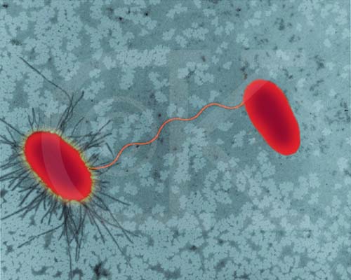
E. coli strains undergoing conjugation (TEM x27,700). This image is copyright Dennis Kunkel http://www.pbrc.hawaii.edu/~kunkel/gallery, used with permission.
Endospores are a method os survival, not one of reproduction. Certain bacteria will form a spore within their cell membrane (an endospore) that allows them to wait out deteriorating environmental conditions. Certain disease causing bacteria (such as the one that causes the disease Anthrax) can be virulent (capable of causing an infection) 1300 years after forming their endospore!
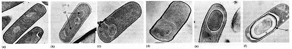
The above image is from: http://www.bact.wisc.edu/Bact303/MajorGroupsOfProkaryotes. Note, the sequence illustrated here goes from left to right.
Eubacteria (eu = true) are the majority of bacteria and are subdivided by their method of energy acquisition into chemosynthetic, photosynthetic, and heterotrophic.
Chemosynthetic bacteria are autotrophic, and obtain energy from the oxidation of inorganic compounds such as ammonia, nitrite (to nitrate), or sulfur (to sulfate).
Photosynthetic bacteria carry out conversion of sunlight energy into carbohydrate energy. Cyanobacteria are the major group of photosynthetic bacteria. Some early cyanobacteria may have formed the oxygen released into the early atmosphere. In addition to chlorophyll a, cyanobacteria also have the blue pigment phycocyanin and the red pigment phycoerythrin.
Filamentous Cyanobacterium, Anabaena sp. (SEM x5,000). This image is copyright Dennis Kunkel http://www.pbrc.hawaii.edu/~kunkel/gallery, used with permission.
Members of this large and diverse group must derive their energy from another organism by feeding. Two main types: saprophytic and symbiotic. Saprophytes feed on dead or decaying material and are important nutrient recyclers. Symbiotic bacteria live within a host multicellular organism and contribute to the health of the host. Examples include cows and other grazing animals: the bacteria convert cellulose from plant leaves and stems eaten by the animal into glucose for digestion by the animal. Normally cellulose is nondigestible.
Possible symbiosis of bacteria within early eukaryotic cells was a major step in the evolution of eukaryotic cells. In 1980, Lynn Margulis proposed the theory of endosymbiosis to explain the origin of mitochondria and chloroplasts from permanent resident prokaryotes. According to this idea, a larger prokaryote (or perhaps early eukaryote) engulfed or surrounded a smaller prokaryote some 1.5 billion to 700 million years ago.
Steps in endosymbiosis. Image from W.H. Freeman and Sinauer Associates, used by permission.
Instead of digesting the smaller organisms the large one and the smaller one entered into a type of symbiosis known as mutualism, where both organisms benefit and neither is harmed. The larger organism gained excess ATP provided by the "protomitochondrion" and excess sugar provided by the "protochloroplast", while providing a stable environment and the raw materials the endosymbionts required. This is so strong that now eukaryotic cells cannot survive without mitochondria (likewise photosynthetic eukaryotes cannot survive without chloroplasts), and the endosymbionts cannot survive outside their hosts. Nearly all eukaryotes have mitochondria. Mitochondrial division is remarkably similar to the prokaryotic methods that will be studied later in this course. A summary of the theory is available by clicking here.
Many heterotrophic bacteria also cause diseases such as strep throat, rheumatic fever, cholera, gonorrhea, syphilis, and toxic shock syndrome. Bacteria can cause disease by destroying cells, releasing toxins, contaminating food, or by the reaction of the body to the infecting bacteria. Bacterial infections can be controlled by vaccinations and antibiotic treatments. Antibiotics interfere with some aspect of the replication of bacteria, and are produced by microorganisms such as fungi, that compete with bacteria for resources. Penicillin, the first antibiotic discovered, inhibits the synthesis of new cell walls in certain types of bacteria. However, the overuse of antibiotics during the past fifty years has led to natural selection favoring antibiotic resistance. There are reportedly more than 50 strains of antibiotic resistant bacteria, necessitating the development of new antibiotics and the frequent change of antibiotics in treatment.
Archaebacteria (now more commonly referred to as the archaea) are considered the oldest and most primitive organisms known. They have significant differences in their cell walls and biochemistry when compared to bacteria. Many scientists propose placing archeans into a separate kingdom, the Archaea.
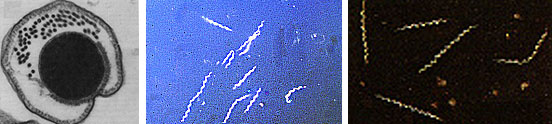
Three types of archaebacteria: methanogens, halophiles, and thermacidophiles. They live in extreme habitats.
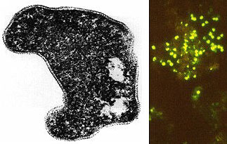
The image above is from http://www.bact.wisc.edu/Bact303/MajorGroupsOfProkaryotes. Sulfolobus acidocaldarius , an extreme thermophile occurs in geothermally-heated acid springs, mud pots and surface soils; it can withstand temperatures from 60 to 95 degrees C, and a pH of 1 to 5. Left: Electron micrograph of a thin section (X85,000); Right: Fluorescent photomicrograph of cells attached to a sulfur crystal.
Fossil evidence supports the origins of life on earth earlier than 3.5 billion years (in scientific nomenclature a billion is referred to as a giga-, abbreviated Ga for giga annum) ago. The North Pole fossils from Australia are such that even more primitive cells must have existed earlier. From rocks of the Ishua Super Group in Greenland come possibly the earliest cells, 3.8 (?) Ga. J. William Schopf of UCLA discovered possibly photosynthetic prokaryotes from North America in 3.5 Ga old rocks, suggesting the existence of even older heterotrophic forms. The oldest known rocks on earth are 3.96 Ga and are from Arctic Canada. Thus, life appears to have begun soon after the cooling of the earth and formation of the atmosphere and oceans.
These ancient fossils occur in marine rocks, such as limestones and sandstones, that formed in ancient oceans. The organisms living today that are most similar to ancient life forms are the archaebacteria. This group is today restricted to marginal environments. Recent discoveries of bacteria at mid-ocean ridges add yet another possible origin for life: at these mid-ocean ridges where heat and molten rock rise to the earth's surface.
Email: mj.farabee@emcmail.maricopa.edu![]()
Last modified:
2000/01/08:17:17:33
The URL of this page is:
gened.emc.maricopa.edu/bio/BIO181/BIOBK/BioBookDiversity_2.html