
Asexual Reproduction | Sexual Reproduction | Human Reproduction and Development
Sexual Responses | Sexually Transmitted Diseases | Reproduction: New and Improved?
Fertilization and Cleavage | Gastrulation | Pattern Formation and Induction | Human Development
The ability to reproduce is one of the unifying characteristics of all living things. Sexual reproduction produces offspring that are genetically different from their parents. Asexual reproduction produces offspring genetically identical to their parent.
Fission, budding, fragmentation, and the formation of rhizomes and stolons are some of the mechanisms that allow organisms to reproduce asexually. The hydra produces buds; starfish can regenerate an entire body from a fragment of the original body. Asexual reproduction allows an organism to rapidly produce many offspring without the time and resources committed to courtship, finding a mate, and mating. The lack of genetic variability in asexually reproducing populations can be detrimental when environmental conditions (for which all the clones are so well adapted) change quickly.
In sexual reproduction new individuals are produced by the fusion of haploid gametes to form a diploid zygote. Sperm are male gametes, ova (ovum singular) are female gametes. Meiosis produces cells that are genetically distinct from each other; fertilization is the fusion of two such distinctive cells that produces a unique new combination of alleles, thus increasing variation on which natural selection can operate.
Rotifers will reproduce asexually when conditions are favorable by having females produce eggs by mitosis. When conditions deteriorate, rotifers will reproduce sexually and encase their zygotes inside a resistant shell. Once conditions improve, these eggs hatch into diploid individuals. Rotifers thus use sexual reproduction as way to survive a deteriorating environment.
Sexual reproduction offers the benefit of generating genetic variation among offspring, which enhances the chances of the population's survival. Costs of this process include the need for two individuals to mate, courtship rituals, as well as a number of basic mechanisms described later.
Human reproduction employs internal fertilization, and depends on the integrated action of hormones, the nervous system, and the reproductive system. Gonads are sex organs that produce gametes. Male gonads are the testes, which produce sperm and male sex hormones. Female gonads are the ovaries, which produce eggs (ova) and female sex hormones.
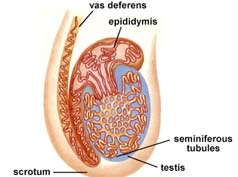
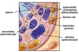
Testes are suspended outside the abdominal cavity by the scrotum, a pouch of skin that keeps the testes close or far from the body at an optimal temperature for sperm development. Seminiferous tubules are inside each testis, and are where sperm are produced by meiosis. About 250 meters (850 feet) of tubules are packed into each testis. Spermatocytes inside the tubules divide by meiosis to produce spermatids that in turn develop into mature sperm.
Images from W.H. Freeman and Sinauer Associates, used by permission.
Sperm production begins at puberty at continues throughout life, with several hundred million sperm being produced each day. Once sperm form they move into the epididymis, where they mature and are stored.
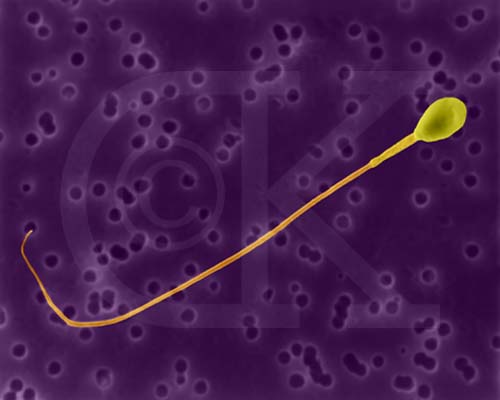
Human Sperm (SEM x5,785). This image is copyright Dennis Kunkel http://www.pbrc.hawaii.edu/~kunkel/gallery, used with permission.
The anterior pituitary produces follicle-stimulating hormone (FSH) and luteinizing hormone (LH). Action of LH is controlled by the gonadotropin-releasing hormone (GnRH). LH stimulates cells in the seminiferous tubules to secrete testosterone, which has a role in sperm production and developing male secondary sex characteristics. FSH acts on cells to help in sperm maturation. Negative feedback by testosterone controls the actions of GnRH.
Sperm pass through the vas deferens and connect to a short ejaculatory duct that connects to the urethra. The urethra passes through the penis and opens to the outside. Secretions from the seminal vesicles add fructose and prostaglandins to sperm as they pass. The prostate gland secretes a milky alkaline fluid. The bulbourethral gland secretes a mucus-like fluid that provides lubrication for intercourse. Sperm and secretions make up semen.
The above images are from http://www.biosci.uga.edu/almanac/bio_103/notes/apr_4.html.
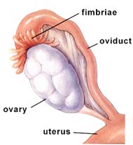
The female gonads, ovaries, are located within the lower abdominal cavity.
Images from W.H. Freeman and Sinauer Associates, used by permission.
The ovary contains many follicles composed of a developing egg surrounded by an outer layer of follicle cells. Each egg begins oogenesis as a primary oocyte. At birth each female carries a lifetime supply of developing oocytes, each of which is in Prophase I. A developing egg (secondary oocyte) is released each month from puberty until menopause, a total of 400-500 eggs.
The above image is from http://www.grad.ttuhsc.edu/courses/histo/notes/female.html.
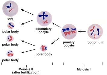
The above diagram of oogenesis is from http://www.biosci.uga.edu/almanac/bio_103/notes/apr_8.html.
After puberty the ovary cycles between a follicular phase (maturing follicles) and a luteal phase (presence of the corpus luteum). These cyclic phases are interrupted only by pregnancy and continue until menopause, when reproductive capability ends. The ovarian cycle lasts usually 28 days. During the first phase, the oocyte matures within a follicle. At midpoint of the cycle, the oocyte is released from the ovary in a process known as ovulation. Following ovulation the follicle forms a corpus luteum which synthesizes and prepares hormones to prepare the uterus for pregnancy.
The secondary oocyte passes into the oviduct (fallopian tube or uterine tube). The oviduct is connected to the uterus.
The above image is from http://www.biosci.uga.edu/almanac/bio_103/notes/apr_8.html.
The uterus has an inner layer, the endometrium, in which a fertilized egg implants. At the lower end of the uterus the cervix connects the uterus to the vagina. The vagina receives the penis during intercourse and serves as the birth canal.
The above image is from http://www.bhs.berkeley.k12.ca.us/departments/science/anatomy/cat/Repro%20system/Female%20Repro.%20organ%20pg.
The female external genitals are collectively known as the vulva. The labia minora is a thin membrane of folded skin just outside the vaginal opening. The labia majora cover and protect the genital area. A clitoris, important in arousal, is a short shaft with a sensitive tip covered by a fold of skin.
The ovarian cycle is hormonally regulated in two phases. The follicle secretes estrogen before ovulation; the corpus luteum secretes both estrogen and progesterone after ovulation. Hormones from the hypothalamus and anterior pituitary control the ovarian cycle. The ovarian cycle covers events in the ovary; the menstrual cycle occurs in the uterus.
Image from W.H. Freeman and Sinauer Associates, used by permission.
Menstrual cycles vary from between 15 and 31 days. The first day of the cycle is the first day of blood flow (day 0) known as menstruation. During menstruation the uterine lining is broken down and shed as menstrual flow. FSH and LH are secreted on day 0, beginning both the menstrual cycle and the ovarian cycle. Both FSH and LH stimulate the maturation of a single follicle in one of the ovaries and the secretion of estrogen. Rising levels of estrogen in the blood trigger secretion of LH, which stimulates follicle maturation and ovulation (day 14, or midcycle). LH stimulates the remaining follicle cells to form the corpus luteum, which produces both estrogen and progesterone. Click here to view a "movie" of the hormone sequences.
Estrogen and progesterone stimulate the development of the endometrium and preparation of the uterine inner lining for implantation of a zygote. If pregnancy does not occur, the drop in FSH and LH cause the corpus luteum to disintegrate. The drop in hormones also causes the sloughing off of the inner lining of the uterus by a series of muscle contractions of the uterus.
The above image is from http://www.grad.ttuhsc.edu/courses/histo/notes/female.html.
Humans do not have a mating season , females are sexually receptive to the male at all times of the year. There are four stages in mating: arousal, plateau, orgasm, and resolution.
During male arousal, blood flows into the three shafts of spongy erectile tissue inside the penis, causing it to become elongated and erect. The female arousal has the swelling of the areas around the vagina, erection of the clitoris and nipples, and secretion of lubricating fluids in the vagina.
After insertion of the penis into the vagina, pelvic thrusts by both partners stimulate sensory receptors in the penis, vaginal walls, and clitoris. The sperm leave the epididymis and secretions of glands form the semen. Orgasm involves contractions of muscles of the penis (male) or vagina (female) and waves of pleasurable sensations.
Resolution reverses the previous phases: muscles relax, breathing slows, the penis returns to its normal size.
Sexually transmitted diseases (STDs) cause over $7 billion to be expended for treatment. STDs can affect the sex partners, fetus, and newborn infants. STDs are grouped into three categories.
STDs that produce inflammation of the urethra, epididymis, cervix, or oviducts. Gonorrhea and chlamydia are the most common STDs in this category. Both diseases can be treated and cured with antibiotics, once diagnosed.
STDs that produce sores on the external genitals. Genital herpes is the most common disease in this class, affecting more than 25 million individuals in the US. Symptoms of herpes can be treated by antiviral drugs, but the infection cannot be cured. Syphilis is a bacterially caused infection, and can, if left untreated, cause serious symptoms and death. However, the disease is curable with antibiotics.
This class of STDs includes viral diseases that affect organ systems other than those of the reproductive system. AIDS and hepatitis B are in this category. Both can be spread by sexual contact or blood. Infectious individuals may appear symptom-free for years after infection.
New techniques have been developed to enhance or reduce the chances of conception. Social conventions and governing laws have developed far slower than this new technology, leading to controversy about moral, ethical, and legal grounds for the uses of such technologies.
The separation of intercourse from pregnancy uses methods blocking one of the three stages of reproduction"
Various contraceptive methods have been developed; none of which is 100% successful at preventing pregnancy or the transmission of STDs. Abstinence is the only completely effective method.
Physical prevention (most effective) include vasectomy and tubal ligation. Vasectomy: the vas deferens connecting the testes with the urethra is cut and sealed to prevent the transport of sperm. Tubal ligation: the oviduct is cut and ends tied off to prevent eggs from reaching the uterus.
Image from W.H. Freeman and Sinauer Associates, used by permission.
Oral contraceptives (birth control pills) usually contain a combination of hormones that prevent release of FSH and LH, inhibiting development of the follicle so that no oocytes are released. Time-release capsules (Norplant) can be implanted under the skin and offer long-term suppression of ovulation. RU-486, the so-called morning after pill, interferes with implantation of the blastula into the uterine wall. Its use as a contraceptive is very controversial.
Barrier methods employ physical (condom, diaphragm) or chemical (spermacides) means to separate the sperm from the egg. Male condoms are fitted over the erect penis; female condoms are placed inside the vagina. Only latex condoms prevent the spread of STDs. Diaphragms cap the cervix and block passage of the sperm into the uterus. Spermicidal jellies or foams kill sperm on contact and must be placed in the vagina prior to intercourse.
About 1 in 6 couples is infertile due to physical or physiological conditions preventing gamete production, implantation, or fertilization. Blocked oviducts (often from untreated STDs) are the leading cause of infertility in females. Low sperm count, low motility, or blocked ducts are common causes of male infertility.
Hormone therapy can cause increased egg production. Surgery can open blocked ducts. About 40 of the cases are due to male problems, 40 due to female problems and the remaining 20% are caused by some unknown agent(s). In vitro fertilization (test-tube babies) is a widely used technique to aid infertile couples.
Fertilization has three functions:
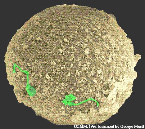
The above image is modified from http://130.102.208.100/FMRes/FMPro?-db=images.fp3&key=32931&-img. Sperm are color enhanced (green) while the egg is color enhanced to gold.
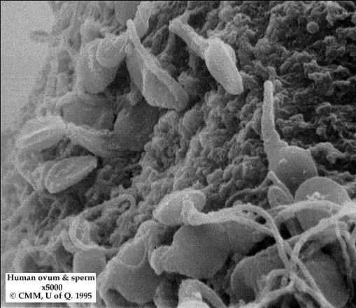
This image is from http://130.102.208.100/FMRes/FMPro?-db=images.fp3&key=32932&-img.
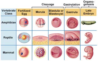
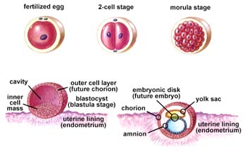
Cleavage is the first step in development of ALL multicelled organisms. Cleavage converts a single-celled zygote into a multicelled embryo by mitosis. Usually, the zygotic cytoplasm is divided among the newly formed cells. Frog embryos divide to produce 37,000 cells in a little over 40 hours.
The blastula is produced by mitosis of the zygote, and is a ball of cells surrounding a fluid-filled cavity (the blastocoel). The decreasing size of cells increases their surface to volume ratio, allowing for more efficient oxygen exchange between cells and their environment. RNA and information carrying molecules are distributed to various parts of the blastula, and this molecular differentiation sets the stage for the layering of the body in the next phases of development.
The above image is from http://www.biosci.uga.edu/almanac/bio_103/notes/apr_11.html.
Image from W.H. Freeman and Sinauer Associates, used by permission.
Gastrulation involves a series of cell migrations to positions where they will form the three primary cell layers.
Ectoderm forms tissues associated with outer layers: skin, hair, sweat glands, epithelium. The brain and nervous system also develop from the ectoderm.
The mesoderm forms structures associated with movement and support: body muscles, cartilage, bone, blood, and all other connective tissues. Reproductive system organs and kidneys form from mesoderm.
The endoderm forms tissues and organs associated with the digestive and respiratory systems. Many endocrine structures, such as the thyroid and parathyroid glands, are formed by the endoderm. The liver, pancreas, and gall bladder arise from endoderm.
Immediately after gastrulation, the body axis of the embryo begins to appear. Chordates have the cells that will form the nervous system fold into a neural tube (which will eventually form the spinal cord). The mesoderm forms the notochord (which will eventually form the vertebrae). The mesoderm at this time forms somites, which form segmented body parts, such as the muscles of the body wall.

Images from W.H. Freeman and Sinauer Associates, used by permission.
Blastulation and gastrulation establish the main body axis. Organ formation occurs in the next stage of the development of the embryo. During organ formation, cell division is accomplished by migration and aggregation.
Pattern formation is the result of cells "sensing" their position in the embryo relative to other cells and to form structures appropriate to that position. Gradients of informational molecules within the embryo have been suggested to provide the positional information to cells. Homeobox genes are pattern genes; they coordinate with gradients of information molecules to establish the body plan and development of organs.
Induction is the process in which one cell or tissue type affects the developmental fate of another cell or tissue. As a cell begins to form certain structures, certain genes are turned on, others are turned off. Induction affects patterns of gene expression through physical contact or chemical signals. Formation of the vertebrate eye is a well known example.
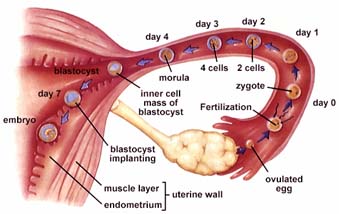
Fertilization, the fusion of the sperm and egg, usually occurs in the upper third of the oviduct. Thirty minutes after ejaculation, sperm are present in the oviduct, having traveled from the vagina through the uterus and into the oviduct. Sperm traverse this distance by the beating of their flagellum. Of the several hundred million sperm released in the ejaculation, only a few thousand reach the egg.
Only one sperm will fertilize the egg. One sperm fuses with receptors on the surface of the secondary oocyte, triggering a series of chemical changes in the outer oocyte membrane that prevent any other sperm from entering the oocyte. The entry of the sperm initiates Meiosis II in the oocyte. Fusion of the egg and sperm nuclei forms the diploid zygote.
Cleavage of the zygote begins while it is still in the oviduct, producing a solid ball of cells (morula). The morula enters the uterus, continuing to divide and becomes a blastocyst.
The above image is from http://www.biosci.uga.edu/almanac/bio_103/notes/apr_11.html.
The uterine lining becomes enlarged and prepared for implantation of the embryo in the trophoblast layer. Twelve days after fertilization, the trophoblast has formed a two-layered chorion. Human chorionic gonadotropin (hCG) is secreted by the chorion, and prolongs the life of the corpus luteum until the placenta begins to secrete estrogen and progesterone. Home pregnancy tests work by detecting elevated hCG levels in the woman's urine.
Maternal and embryonic structures interlock to form the placenta, the nourishing boundary between the mother's and embryo's systems. The umbilical cord extends from the placenta to the embryo, and transports food to and wastes from the embryo.
The above image is from http://www.biosci.uga.edu/almanac/bio_103/notes/apr_15.html.
The period of time from fertilization to birth (usually 9 months) is divided into trimesters, each about three months long. During pregnancy the zygote undergoes 40 to 44 rounds of mitosis, producing an infant containing trillions of specialized cells organized into tissues and organs.
The three embryonic tissue layers form. Cellular differentiation begins to form organs during the third week. After one month the embryo is 5 mm long and composed mostly of paired somite segments. During the second month most of the major organ systems form, limb buds develop. The embryo becomes a fetus by the seventh week. Beginning the eighth week, the sexually neutral fetus activates gene pathways for sex determination, forming testes in XY fetuses and ovaries in XX fetuses. External genitalia develop.
The fetus increases in size during this trimester, and bony parts of the skeleton begin to form. Fetal movements can be felt by the mother.
During this trimester the fetus increases in size. Circulatory and respiratory systems mature in preparation for air breathing. Fetal growth during this time uses large parts of its mother's protein and calcium intake. Maternal antibodies pass to the fetus during the last month, conferring temporary immunity.
Birth is a positive feedback hormonal mechanism. During birth the cervix dilates to allow passage of the fetus. Uterine contractions propel the fetus through the birth canal, usually head first. Hormonal control of the birth process involves the release of oxytocin and prostaglandins, which are stimulated by uterine contractions, which stimulate more hormones that cause more contractions....etc.
The first stage of birth lasts from beginning of contractions to the full (10 cm) dilation of the cervix. Membranes of the amniotic fluid rupture, lubricating the vagina.
Strong uterine contractions of a minute in duration separated by two to three minute intervals propel the fetus down the birth canal. Abdominal muscles relax in synchrony with the uterine contractions.
After delivery of the baby, the umbilical cord is clipped and cut. The placenta (or afterbirth) in expelled through the vagina.
Nursing mothers have their hormone levels and uterine size return to normal much faster than non-nursing mothers. Breasts develop the capability for milk secretion about the mid point of pregnancy. Secretion of milk does not occur until delivery, and the action of prolactin. Suckling by the infant causes production of oxytocin to promote release of milk into the ducts emptying into the nipple.
The stages of life are noted by the acquisition of social, physical, and mental skills. Other signs of maturation include tattoos, body piercing, motorcycles, and backward-turned baseball caps.
Infancy lasts from birth until age two. Infant reflexes concern finding the nipple: suckling can occur minutes after birth, and the rooting reflex is a tactile clue for the infant to turn its head toward a touch. During the first year the infant passes through a series of developmental stages.
Childhood begins at two and lasts until puberty. Physical growth is accompanied by increasing development of motor and language skills. Play, make-believe, and increases in memory and attention span also occur.
Growth accelerates during puberty. Boys increase in height by 3-5 inches per year, girls 2-4 inches. The heart doubles in size, and hormones stimulate sexual maturity. Growth ends during the end of adolescence, and aging starts during the late twenties. Life expectancy (47 and 50 years for males and females in 1900; 74 and 81 in 1996) has increased.
Email: mj.farabee@emcmail.maricopa.edu![]()
Last modified:
2000/01/09:10:56:45
The URL of this page is:
gened.emc.maricopa.edu/bio/BIO181/BIOBK/BioBookREPROD.html