
UNIVERSITY OF THE WEST INDIES
BL14A - BIODIVERSITY I : THE PLANT KINGDOM
PRACTICAL 3 : PTERIDOPHYTESTHIS PRACTICAL REQUIRES YOU TO SPEND A FEW MINUTES IN THE LAB SESSIONS OF THE PREVIOUS TWO WEEKS SETTING UP CULTURES & CHANGING SOLUTIONS.
A. THE FERNS
I. Fern Gametophytes
Week 1
During week 1, each pair of students will set up a fern gametophyte culture for
examination in week 3. Pour 10-15 ml
of the nutrient solution into a petri dish. (This solution simply contains inorganic salts
and fungicide). Take the fern frond provided and scrape some spores from the sori onto the
surface of the medium of the petri dish.
Label your petri dish with your name and date.
Another dish has been set up much earlier and older gametophytes from this should be available during the laboratory session.
Week 3
Use a pasteur pipette to transfer a drop of culture medium containing the gametophytes
to a slide.
Examine and draw the 1 week old gametophytes. An older heart-shaped stage may also be
available.
 1-2 week
old fern gametophyte
1-2 week
old fern gametophyte  Older heart-shaped stage
II. Fern Sporophytes
(i) Examine in some detail the commonly-cultivated Sword Fern, Nephrolepis sp.
Draw a diagram to show the basic morphology of this sporophyte
(ii) Mount some sporangia from this fern in water on a microscope slide.
Having looked at these under the microscope,
irrigate the slide with 90% ethanol, watching the sporangia all
the while.
Record in words and diagrams what you see.  This is what a fern
sporangium looks like.
This is what a fern
sporangium looks like.
(iii) A demonstration of the diversity of ferns is located on the side bench.
Familiarise yourself with these plants.
B. THE FERN ALIES
I. Club & Spike mosses
Club mosses grow wild in the wetter parts of Barbados.
They are, nonetheless, a relic group of plants, most being extinct.
To examine the strobili of these plants we will use a whole mount clearing technique.
Clearing methods transform thick masses of tissue into translucent tissue through chemical
treatment.
By infiltrating the tissue with a fluid, the medium of the tissue is now of uniform
refractive index allowing
light rays to pass through with minimal scattering or absorbance by pigments.
Week 1
Place strobili of Lycopodium and Selaginella in fixative FPA (formalin: propionic acid: 50% ethanol; 5:5:90).
Week 2
Transfer the strobili to 70% ethanol.
Week 3
Transfer the strobili to clearing fluid for 30 minutes and then mount in the same
solution on a cavity slide
(clearing fluid = lactic acid: chloral hydrate: phenol: clove oil: xylene: benzyl benzoate
- 2:2:2:2:1:1).
Examine under low power. There should be a difference in spore size for one of the plants.
If you cannot see the difference consult a Demonstrator.
You do not need to prepare diagrams of these cones.
Draw a diagram of Lycopodium cernuum to show the main features of
its morphology.
Draw a diagram from the prepared slide of the Lycopodium strobilus.
Note that all the spores are of the same size.
 |
 |
|---|---|
| Lycopodium with cones | L.S. Lycopodium cone |
Two species of Selaginella are provided, S. serpens and S.
plana. Both of these are found in Barbados.
Draw a diagram to show the morphology of one of these.
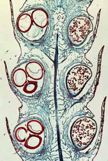
Draw a diagram from a prepared slide of the Selaginella strobilus.
Note the dissimilarly-sized microspores and megaspores and the ligule on each sporophyll
(absent from Lycopodium and in the micrograph below only visible on the bottom
right sporophyll).
 |
 |
|---|---|
| Selaginella serpens | L.S. Selaginella cone |
II. Horsetails & Whisk Ferns
Only one genus - Equisetum - of this once successful group, the
horsetails, still survives. Take a good look at the preserved Equisetum
sporophytes, noting the apical strobilus and the rings of small leaves at the nodes.
DO NOT DRAW THESE.
 |
This is a fertile shoot tip of Equisetum.
Be able to recognise this group, the horsetails, but we will not study them in this course. |
| The whisk ferns (Psilophytes) have no roots and usually no leaves. Instead of roots they have rhizomes with absorptive rhizoids. Psilotum nudum is found in the Caribbean and exemplifies this group. Do not draw this plant but examine the preserved specimen, noting its simple form. | 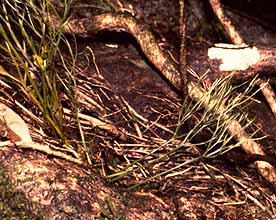 |
TO BE SUBMITTED:
Fern gamephytes:
(i) Diagrams of 1 week old gametophyte
(ii) Diagram of older stage if available
Fern sporophytes:
(i) Annotated diagram of spore liberation
(ii) Diagram of gross morphology of Nephrolepis
Fern allies:
(i) Diagram of L.S. Lycopodium strobilus
(ii) Diagram of L.S. Selaginella strobilus
(iii) Diagram of gross morphology of Lycopodium cernuum
(iv) Diagram of gross morphology of Selaginella sp.
![]()