|
|
Quiz Me! |
Microscopy. Procaryote anatomy: membranes & internal structures. |
|
|
|
Lecture Index | ||
|
|
Course Resources page |
Last revised: Friday, January 7, 2000
Reading: Ch. 2; Ch. 3 (p. 37-51) in Prescott et al, Microbiology, 4th Ed.Note: These notes are provided as a guide to topics the instructor hopes to cover during lecture. Actual coverage will always differ somewhat from what is printed here. These notes are not a substitute for the actual lecture!Copyright 2000. Thomas M. Terry
Microscopy
Units of Measurement
- centi- (10-2)
- milli- (10-3)
- micro- (10-6)
- nano- (10-9)
- Ångstrom (10-8 cm., 0.1 nm)
- I'd like to review dimensions before I proceed
Size and Scale
- View "How Big?" from Cells Alive
- View Hyperspace tour. (Note: you need a password to the Biology Place to visit this site. Anyone can apply for a week's free pass to all Biology Place materials. Subscription rates are very reasonable, both for students and faculty. This is one of the best resources for biology on the Web.)
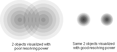
Resolving Power
- measures the ability to distinguish small objects close together
              	        0.5 (lambda)
             r.p. =   ______________
                       (N sinØ)
where lambda = wavelength of illuminating light.

- for light scope, can improve R.P. by making lambda smaller or sinØ larger.
- R.P. is smallest for violet light, but because human eye is more sensitive to blue, optimal R.P. is achieved with blue light (~450 nm). Use filters to remove other light in best microscopes
- n sinØ is called numerical aperture. It measures how much light cone spreads out between condenser & specimen. More spread = better resolution. Ø = angle of light cone; maximum value is 1.0
- n = refractive index. n = 1.0 in air. Can --> with certain oils (up to 1.4),called immersion oil.
note: N.A. is property of lens. Look on side of lens to see- Theoretical limit of R.P. for light scope is 0.2 Ám.
Optical Instrument Resolving Power RP in Angstroms Human eye 0.2 millimeters (mm) 2,000,000 A Light microscope 0.20 micrometers (Ám) 2000 A Scanning electron microscope (SEM) 5-10 nanometers (nm) 50-100 A Transmission electron microscope (TEM) 0.5 nanometers (nm) 5 A Light Microscopes
- Bright-Field Microscope

- Advantages: convenient, relatively inexpensive, available
- Disadvantages: resolving power 0.2 micrometers at best, can recognize cells but not fine details
- Needs contrast; cells are mainly water. Easiest way to view cells is to fix and stain.
- Fixation
- preserves cells; disrupts proteins, prevents decay/degradation.
- typical treatments: heat, formalin, glutaraldehyde
- Staining
- Simple Stains
- adds colored compounds --> contrast
- basic dyes: e.g. methylene blue, crystal violet. Cations (have + charges)----bind to - charge groups on proteins, nucleic acids
- acidic dyes: e.g. eosin, acid fuchsin. Anions (have - charges); bind to + charges on proteins, phospholipids
- Differential Stains
- allow differentiation between different organsisms. Examples:
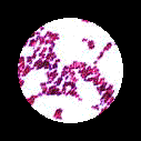
- Gram stain----lab this week. Distinguishes 2 major groups of bacteria: + and -.
- Spore stain----lab next week. Malachite green binds specifically to compounds in endospore wall.
- Acid fast stain----not done in 229 lab. Allows detection of some bacteria with waxy coat (Mycobacteria).
- Optical path: source, condenser lens, specimen, objective lens, eyepiece lens
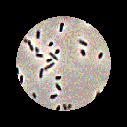
- Phase-Contrast Microscope

- Cells are mostly water, very little contrast from surrounding medium, so not very visible in light
- Phase scope converts slight diffs. in refractive index and cell density into variations.
- Scope uses annular stop below condenser: thin transparent ring in opaque disk --> hollow light cone. As light passes through specimen, some rays are deviated and retarded by 1/4 wavelength.
- Have phase plate in objective lens: transparent optical disk with phase ring.
- Undeviated light passes through ring, is advanced by 1/4 wavelength --> bright background. But deviated light doesn't pass through phase ring, so is not advanced. When light gets focused, deviated rays cancel out with undeviated rays, producing dark image where objects were.
- Advantage: can see live material w/o staining
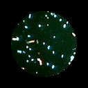
- Fluorescence Microscope

- Fluors are chemicals that adsorb light to produce excited electrons, later reradiate light = flourescence.
- To use, need special type of microscope. Must illuminate with ultraviolet or violet light (--> excited fluor). Need filters to remove this light from light traveling to ocular lens; only fluoresced light emitted from object will then appear to eye. Need dark field condenser to create dark background.
- Can couple flour to specific probe molecules (usually antibodies), bind to preparation. If sample is illuminated with wavelength of exciting light, then filter out that wavelength to prevent reaching sample, see nothing. But if fluorescence occurs, diff. wavelength light produced, see object.
- good technique to detect specific microbe in complex sample. (e.g. detect gonococcus in vaginal smear). Requires correct microscope, fluors, technical skill.
- For further information, see "Cell structure and Microscopy"
Electron Microscope
- Physicists discovered electrons have wave properties. Can use magnetic coils like lenses to focus beams of electrons. Basic design of EM similar to light scope
- But: electrons don't scatter from H, C, O, N: must add heavy atoms (e.g. Pb, Ur, Os, Gold) as stains.
- Also, electrons are scattered by air molecules. So must remove air from microscope with vaccum pump. But water in specimen will evaporate, so must be removed by dehydration after fixation. Cannot view living specimens.
Transmission Electron Microscope (TEM)
- see slide. R.P. approx. 1000x better than light; 0.2 nm, instead of 0.2 micrometers.
- excellent for seeing internal detail. But cannot use with large/thick specimens.
- View TEM of E. coli
Specimen Preparation
- specimen must be thin. Use grids with thin film supports. Prepare thick materials by sectioning with glass knives --> sections about 20-100 nm thick. Prepare small preparations (viruses, or subcellular particles) by negative staining. Alternatively, use metal shadowing.
- Freeze-etch procedure. Freeze specimen, fracture --> follows membrane contours. Etch in vacuum (water sublimates), then shadowcast with metal --> replica film. Dissolve specimen, view film.
Scanning Electron Microscope (SEM)
- Same principle as TV screen, except reflected (secondary) electrons used to produce magnified image.
- complementary to TEM. Only see surface view --no internal detail visible. Infinite depth of focus, in contrast to light scopes.
- R.P. around 2 nm at best, usually a bit poorer. (100x better than light scope, not as powerful as TEM)
- View SEM of E. coli
Specimen Preparation
- easy! Fix & dry specimen. Shadow with thin metal film (e.g. gold).
- Mount on block and scan. (Note: sometimes possible to use ordinary air-dried material; but charge builds up on surface, distorts image).
Prokaryote Anatomy
Composition of a bacterial cell
- Important points:
- Water makes up ~ 70%
- Macromolecules make up ~ 26% ( ~90% of dry wt)
- The most abundant molecules other than water are proteins (~50% of dry wt)
- Small molecules & ions make up 4% (~10% of dry wt)
How big are bacteria?
- View graphic of bacterial sizes
- Smallest bacteria are Mycoplasmas, as small as 0.2 micrometers (almost as small as largest poxviruses!)
- Accepted wisdom is that bacteria are smaller than eukaryotes. But certain cyanobacteria are quite large; Oscillatoria cells are 7 micrometers diam, size of red blood cells. And certain eukarotes (e.g. Nanochlorum eukaryotum) are very small, only 1 to 2 micrometers, but true eukaryotes (nucleus, chloroplast, mitochondrion are present). So size difference is, like many generalizations, only a useful yardstick, not an absolute truth.
- Epulopiscium fishelsoni, discovered in 1985 in intestinal tract of sturgeonfish, is an enormous, cigar-shaped cell, as large as 80 x 600 micrometers (that's .6 mm, large enough to be seen by the naked eye). Amazingly, this cell is prokaryotic! Initial evidence by EM was hard to believe, but confirmed ty rRNA comparisons with other organisms, a cousin of Gram-positive Clostridium genus.
Basic Structures of Prokaryotic Cells.
Cell membrane
- function: permeability barrier, prevents cell contents from leaking away. Very impermeable to polar & charged molecules (most biomolecules, also small ions such as K+, H+). Only water, lipid-soluble materials pass freely.
- But cells have many specific protein carriers in membrane, carry out selective transport.
Membrane Components and Structure
- Chemical Composition of membranes: ~50% lipid, 50% protein
- Membrane lipids are typically phospholipids, contain two fatty acids joined to glycerol by ester bonds (See Fig. 2.7 in text).
- View interactive pdb file of phospholipid. (Note: to view this file on your computer, the Chime plug-in must be installed in your plug-in folder.)
- Phospholipids assemble into a lipid bilayer.
- Biological membranes contain lipid bilayer + many embedded proteins. Proteins "float" in fluid lipid layer = "fluid mosaic model" of membrane structure.
- Other membrane lipics: sterols and hopanoids, rigid planar molecules, may add strength to membranes. (See Fig. 3.6) Sterols usually absent in bacterial membranes (common in eukaryotic membranes, account for 5-20% of total lipid). But hopanoids present, probably stabilize membranes. Enormous % of fossil fuels = hopanoids (global mass est. at 1011-12 tons, about same as total mass of organic carbon in all living organisms, ~ 1012 tons!)
- Note: Archaea have different type of lipids; not fatty acids, instead built of isoprene units. (See text Fig. 3.19). Typical membrane lipids are called diglycerol diethers or glycerol tetraethers (see Fig. 20.3). The latter molecule produces lipid monolayers instead of bilayers.
- View physiology of Archaea. (Note: you need a password to the Biology Place to visit this site.)
- Membrane proteins contribute stability and many functions, such as transport.
Inside the Cell
Cytoplasm
- largely water (~70%)
- contains as many as 1000 different enzymes, organized into metabolic pathways
- many ribosomes (occupy up to 25% of cell volume, use up to 90% of cell energy), used to make new proteins (mostly enzymes)
- Ribosome size measured in Svedberg (S) units, orginally from ultracentrifugation studies.
- Bacterial ribosomes = 70 S; but eukaryotic ribosomes larger, = 80S
- Ribosomes dissociate into subunits. 70S = 30S + 50S. (prokaryote)
80S = 40S + 60S (eukartyote)Nucleoid
- Bacteria lack nucleus; DNA contained in cytoplasm in form of tighly coiled circular single DNA molecule, called the bacterial chromosome.
- Length is about 1000x longer than cell. Total size for smallest bacteria less then 1,000,000 base pairs. For E. coli (fairly sophisticated bacterium), 4,700 kilo base pairs (kbp).
- Bacterial DNA organized in dense staining region = nucleoid. DNA is supercoiled
- Replication of DNA in procaryotes is fundamentally different from eukaryotes: no mitosis, no cytoskeleton. Instead, DNA division occurs, two copies separated to opposite ends of cell, cell pinches off into two copies.
- Plasmids (small circular DNA elements) are found in virtually all bacterial cells.
Inclusion bodies (found in some, not all, cells)
- stored energy
- poly-beta-hydroxybutyric acid (fatty material, very high energy)
- glycogen (polysaccharide, made of glucose sugare; good energy source)
- polyphosphate (used to store phosphate, often a limiting nutrient)
- sulfur granules (common in some photosynthetic bacteria)
- magnetosomes (common in magnetotactic bacteria); allow cells to orient to magnetic fields
- gas vesicles
- found in many photosynthetic bacteria and cyanobacteria
- involved in flotation: form rigid air-filled sac
- surrounded by rigid protein (not lipid) membrane. Impermeable to liquids, but permeable to gas.
Take a Self-Quiz on this material
Return to Lecture Index
Return to MCB 229 Course Resources page