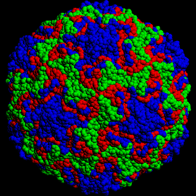
Rhinovirus 14 as colored by its proteins
 MPEG Version (593K)
 Quicktime version (1271K)
Summary
Rhinovirus 14 illustrated with VP1 in blue,
VP2 in green and VP3 in red (VP4 is inside
and not visible).
Each amino acid is represented by a 4 Angstrom sphere.
Rotations have a range of 72 degrees, i.e.
1/5 of a fullrotation,
Rotations along the X axis are every 4 degrees.
Rotations along the Y axis are every 3 degrees.
The version usually distributed is one that has images reduced
by at least 40%(or less) making the movie small enough to
keep on a single HD 3"1/2 disk. With full size images the
movie file would be more than 8MB.....
The compression used is Apple garphics which keeps high quality
of images even with reduced sizes. The compressed video verion
would be only ~300K instead of ~1.2MB but the quality is too low.
Images were calculated on a Silicon Graphics 310 VGX Workstation (SGI) (R3000MIPS
microprocessor) with the program 'srf' (surface Rendering by Foliation) from Dr. Michael Connolly.
For the X directions (19 frames) the calculation took a total of 3 hours on the SGI.
For the Y directions (25 frames) the calculation took a total of 4.5 hours on the SGI.
The default file format was 'SUN' raster with 400x400 pixels, which was converted to the
SGI'rgb' format (SGI utility 'fromsun') and then to the Macintosh PICT format (SGI utility 'topict').Files were then transfered to a Mac with FTP (file transfer protocol, binary) and the type and creator changed with 'FileTyper' to have the correct PICT format , and given a
creator of TeachText).
 Go to the
top page
Go to the
top page
© 1994 Jean-Yves Sgro. Institute for Molecular Virology/ jsgro@facstaff.wisc.edu