Rhinoviruses
__________________________________________________________________________
(For a more General Reference: Picornaviridae and Their Replication, CHAPTER 20. Roland R. Rueckert. Virology. Second Edition, edited by B.N. Fields, D.M. Knipe et al. Raven Press, Ltd., New-York - 1990)
___________________________________________________________________________
Classification
Rhinovirus is part of the Picornaviridae family. That is the *pico*rna*.
'PICO' means small, 'RNA' signifies that these are single standed positive
sense RNA viruses. Thus these represent small RNA viruses.
The picornavirus family is divided in 5 Genera and further subdivided into members.
- Enterovirus
- Human polioviruses 1-3 (serotypes=3)
- Human hepatitis virus A (serotypes=1)
- Theiler's murine encephalomyelitis (serotypes=1)
- Rhinovirus
- Human rhinoviruses 1-100 (serotypes=100)
- Bovine rhinoviruses 1-2 (serotypes=2)
- Aphtovirus
- Foot-and-mouth disease virus 1-7 (serotypes=7)
- Cardiovirus
- Encephalomyocarditis (EMC), Mengo (serotypes=1)
- Unassigned
- Equine rhinoviruses 1-2, Cricket paralysis... (serotypes>3)
Polyproteins
The single RNA is translated into a polyprotein that is subsequently cleaved.
The way the proteins cleave themselves out is slightly different amongst the different genera. For our structural purposes suffice it to say that the coat proteins are
products of the cleavage of the P1 precursor protein.
       <--------  P1 -------->
       ---------------------------------------------------------------
Vpg---|  VP0   |  VP3  |  VP1 |2A| 2B |    2C   |3A|3B|  3C  |    3D  |
       ---------------------------------------------------------------
P1 - > VP0 + VP3 + VP1 and then VP0 -> VP2 + VP4 once the virus has assembled.
Vpg (virion protein genome) is attached to the 5'end of the RNA.
Coat Proteins
In shematics and color graphics and movie representations I use the convention:
- VP1 = blue
- VP2 = green
- VP3 = red
- VP4 = yellow
Coat Protein Mass
_______________________________________
|  VP1	|  VP2	|  VP3	|  VP4	|  P1
32,300	28,500	26,200	7,200	94,200
_______________________________________
VP1-3 have the now famous wedge-like protein fold motif shared by most
virus structures solved by X-ray crystallography. It is created by a
common CORE made up of 8 antiparallel Beta-strands forming a Beta-barrel
structure. Proteins amongst viruses mainly differ in the loop insertions
between the strands. These insersions accounts for a lot of the virus
external appearance and also are the target structures for antibodies in
the case of an animal virus. (see below). Interestingly there is no sequence
conservation in the Beta strands structures of viruses although the structure
is very well conserved.
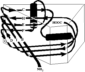
Here is a representation of the 8-stranded Beta barrel. The beta strands
orientation is marked with the sybols: V,^,> and < depending on the position in
the drawing. The Beta strands are labeled BIDG and CHEF. In the first plant
virus solved an A Beta strand is found at the N terminus but is not preserved
in other viruses.The loop names are also marked on the edges (BC,HI,DE,FG,CD,EF,
GH,EF).
The *** represent the 2 Alpha- helices. The helix between loop EF and strand F
is in the back.

The VP4 protein is much shorter and is found only on the interior surface of
the virus. It results from the cleavage of VP0 into VP2 and VP4 which
occurs after assembly. In poliovirus the N-terminus is myristalated.
Icosahedral Capsid
60 of each of the coat proteins assemble into an icosymmetric structure
with icosahedral symmetry. Rhinovirus has a triangulation number of T=1
but is also refered to as a P=3 for Pseudo-T=3 structure. A T=3 structure
occurs when all 180 (3 protein for each of the 60 facets) are chemically
identical but have to assume slightly different neighbouring environment:
The proteins which are around the 5-fold axis have 4 neighbours while the
ones at the 3-fold axis have 5 neighbours.
                      ___________________
                     /\                 /\
                    /   \             /   \
                   /      \         /      \
                  /         \     /         \
                 /            \ /            \  T=1:
                /            - V -            \  A 20 facets
               /         _ /  /|\  \ _         \  Icosahedron.
              /      _ /     / | \     \ _      \   Contains
             /   _ /        / 60  \        \ _   \	5-fold vertices.
            /_ -           /___|___\           - _\ T=3:
            \  - _             |     20      _ -  /	Each of the 20
             \     \ _         |         - /     /	facets can be
              \        \ _     |     - /        /	subdivided into
               \           \ _ ^ _ /           /     60 total triangles.
                \              |              /     Each of these 60
                 \            / \            /     triangles contains
                  \         /     \         /    approximately one
                   \      /         \      /   each of the VP coat proteins.
                    \   /             \   /
                     \/_________________\/
               VP1  |   VP1
                    5   		     Within each of the 60
               .  '/ \` .                     triangles fits about 1 each of
            ,     /   \   ` .                   VP1-3. The symmetry axes are
        ,    VP1 /     \ VP1  ` .      \         labeled 5,2,3. p3 is a pseudo
    ,           /  VP1  \       ` ` .   \         3-fold axis within the triangle.
,              /         \       `    ` .\        Around the icosahedral
              /         / \       ` VP3  '       5-fold are five VP1s.
             /         /   \       `. . ./      But around the 3-fold
            /         /     \     /            are 3 VP2s and 3 VP3s
     VP3   /_______p3/       \   /     /      alternating.
          /         \         \ /              In Plant viruses with T=3
 \       /           \   VP3   \ VP2 /          symmetry the similarity is
        /     VP2     \         \                with VP1=A, VP2=C, VP3=B.
    \  /               \         \  /   VP3
VP2__3/___________2_____\_________\3 __
                 \                 |
      \           \               / \  VP2
   VP2 \  VP3      \    VP2      /    \
        \           \           /  VP3  \
         \           \_________/
          \          /        /
                    /
In T=3 plant viruses each protein is synthezised independently while in
the polioviruses they arise from the cleavage of P1 as illustrated
above. It is worh noting that the VP proteins issued from a single
P1 precursor do not form a triangular face as the previous drawing
might suggest. Instead they form what is usually refered to as the
                             *Biological* asymmetric
                   5         unit as opposed to the *crystallographic*
                  / \         asymmetric unit. An asymmetric unit is
                 /   \         a set of minimum non-redundant information
                /     \        necessary to reconstruct an icosaheron from
               /       \       it using only the icosahedral symmetry.
              /         \       The complete icosahedron is indeed
      _______/   VP1   /       mathematically obtained wether one uses
     /      /         /       a triangular asymmetric unit or this
    /      /         /        biological unit in which the VP3 area has only
   /      /_______  /        been switched to the right!
  / VP3  /         \
 /      /           \
 \     /     VP2     \
  \   /               \
   \3/_________________\
                  2
Canyon and Drug Binding in VP1 Hydrophobic Pocket
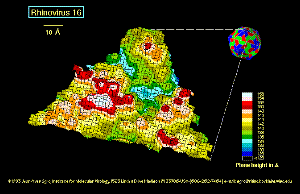
The VP1 proteins form a small cylindrical protrusion at the 5-fold axis.
Around that protrusion is a depression sometimes refered to as the 'canyon'.
This canyon is the receiving site for the cellular receptor which has been
co-crystallized in this position by the crystallographers at Purdue University,
and also observed boud there by cryo-EM reconstruction.
Here is a projection of the canyon onto one of the 60 triangular facets. The
scales to the left shows the position in Angstroms along the X axis and range
from 4 to 42. The bottom scale shows the size in Angstroems for the Y axis
ranging from 01 at the right to 61 at the left; (the axis choice was slected
by the crystallographers). The canyon appears as numbers within a background
of + symbols. Numbers range from 1 to 4, 4 showing the deepest part of the
canyon. The rim of the canyon is arbitrarily set at 137 Angstroems from the
center of the particle. The output was prepared by the program V-suf
(Rossmann and Palmenberg (1988), Virology 164,373-382)

The VP1 protein has also the peculiarity of having a hydrophobic pocket
accessible from the surface via a small 'pore' entrance. It is indicated
by the symbol () in the above drawing (approximate position). In some of the
virus structures resolved by X-ray crystallography a substance can be
found in this hydrophobic cavity. The nature of the compound is not known.
It is refered to as a Sphingolipid in the case of poliovirus and as a sugar
in the case of rhinovirus 1a, but crystallographers only see some 'extra'
electron density. The pharmaceutical company Sterling Winthrop has synthezised
compounds, often refered to as WIN-drugs, that diffuse readily in the VP1
pocket. (see e.g. Heinz et al. J. Virol. (1989) vol.63, pp 2476-2485,
Genetic and Molecular Analyses of Spontaneous Mutants of Human Rhinovirus14
That Are Resistant to an Antiviral Compund). Other companies also study
similar compounds (Jansen, Chalone, Sandoz) but none of them are ready
for commercial use.
WIN 52035
  ___       ____
 |   N     /    \              O____N
 |    \\__<  ()  >-O--  /\  __/    ||
 |___O/    \____/     \/  \/  \\___||__CH3

Oxazoline  Phenoxy   Aliphatic  Isoxazole	Chain
The effect of the compound is to raise the canyon floor and
prevent attachement to the cellular receptor that now cannot fit
properly in the canyon and / or impair the uncoating of the capsid upon cell
entry. The presence of such a compound in the structure confers thermal
stability to the virion.
It is suggested that this is a trigger mechanism that allows the
virus to survive in vivo. For instance poliovirus is stable in
acid conditions and survives inside the stomach while rhinovirus
does not.

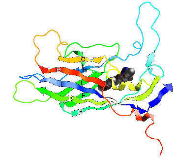
This ribbon diagram shows
the VP1 protein of rhinovirus 14 with the WIN drug bound.
Antigenic Sites
Antigenic sites have been determined by escape mutations
Rueckert et al. (1986), Virus Attachement and Entry Into Cells, American Society for
Microbiology: Location of four neutralization antigens on the three-
dimension; surface of a common-cold picornavirus, human rhinovirus14;
Rossmann et al. (1985), Nature, 317,145-153. Structure of the human
common cold virus and functional relationship to other picornaviruses)
Viruses were resitant to a panel of murine monoclonal antibodies. "Mutations
were clustered in the serotype-variable regions of the amino acid sequences
fo the virus capsid proteins". These antigenic areas are found to protrude
out of the surface of the virus. Refer also to the above diagram, showing
the depth of the canyon...
Four neutralization Immunogens (NIm) sites have been resolved by
cross-neutralization tests. Sites are labeled NIm IA, IB, II, and III. The
** symbols approximate the size of the area. IB is a smaller area than IA.
                   5
                  / \		IA::(VP1): Q83,K85,D138,S139
                 / IA\		  IB::(VP1):D91,E95
                /  ** \
               /       \	     II::(VP2):E136,S158,A159,E161,V162
              /     IB  \		 (VP1):E210
             /         / \
            /         /   \		III::(VP3):N72,R75,E78,G203
           /         /     \		     (VP1):K287
          /____*__  /       \
         /    ** * \         \
        /     II ** \     *   \
       /             \   III*  \	The major antigenic site is on the BC
    3/________________\_________\3	loop in VP1 and VP3 but in the EF loop
                  2			in VP2.
Available 3D Coordinates
Rhinovirus has been crystalized as a native virus but also with
various drugs. The group from Michael Rossmann's at Purdue Univeristy
has released those structures to the protein databank (PDB) at brookhaven
which are available on anonymous ftp (pdb.pdb.bnl.gov [130.199.144.1]):
-------------------------------------------------------------------------------
PDB
ENTRY#	VIRUS					AUTHORS			  DATE
-------------------------------------------------------------------------------
1R1A  RHINOVIRUS 1A                             M.ROSSMANN ET AL.         12/88
4RHV  RHINOVIRUS 14(HUMAN)                      E.ARNOLD,M.ROSSMANN        1/88
2RS1  RHINOVIRUS/ANTIVIRAL AGENT 1S COMPLEX     M.ROSSMANN ET AL.         10/88
2RR1  RHINOVIRUS/ANTIVIRAL AGENT 1R COMPLEX     M.ROSSMANN ET AL.         10/88
2RM2  RHINOVIRUS/ANTIVIRAL AGENT 2 COMPLEX      M.ROSSMANN ET AL.         10/88
2RS3  RHINOVIRUS/ANTIVIRAL AGENT 3S COMPLEX     M.ROSSMANN ET AL.         10/88
2R04  RHINOVIRUS/ANTIVIRAL AGENT 4 COMPLEX      M.ROSSMANN ET AL.         10/88
2RS5  RHINOVIRUS/ANTIVIRAL AGENT 5S COMPLEX     M.ROSSMANN ET AL.         10/88
2R06  RHINOVIRUS/ANTIVIRAL AGENT 6 COMPLEX      M.ROSSMANN ET AL.         10/88
2R07  RHINOVIRUS/ANTIVIRAL AGENT 7 COMPLEX      M.ROSSMANN ET AL.         10/88
1R08  RHINOVIRUS/ANTIVIRAL AGENT 8 COMPLEX      M.ROSSMANN ET AL.         10/88
1R09  RHINOVIRUS 14/R61837                      M.ROSSMANN ET AL.          5/90
1RMU  RHINOVIRUS MUTANT((1)C199Y)               M.ROSSMANN ET AL.         10/88
2RMU  RHINOVIRUS MUTANT((1)V188L)               M.ROSSMANN ET AL.         10/88
1HRI  RHINOVIRUS 14(HUMAN)/ AGENT SCH 38057 	A.ZHANG,R.G.NANNI,E.ARNOLD10/92
-------------------------------------------------------------------------------
2PLV  POLIO VIRUS                               D.FILMAN,J.HOGLE          10/89
2MEV  MENGO VIRUS                               M.ROSSMANN                 4/89
-------------------------------------------------------------------------------
(The PDB entry #'s above are linked to the Molecules 'R Us forms query at NIH,
which will produce an image of any protein that has coordinates in the Protein
Data Bank. Try it!)
Images and Animations of Rhinoviruses
Check out the Rhinomovies page!
Note
A completely ascii version of this document is available for those who can
not view the images.
Published on the Internet. Reproduction granted for educational purposes.
 Go to the
top page
© 1994 Jean-Yves Sgro. Institute for Molecular Virology/ jsgro@facstaff.wisc.edu

