The branch of science that is committed to the study of tissues is called histology. Though with plants the term plant anatomy is heard just as often. The term tissue stems originally from a misinterpretation. N. GREW coined it in 1682 since he thought that the scaffolding of the cell wall would consist of very fine threads and that the organization of the plant would resemble the layering of a larger number of Brussels lace on top of one another. He spoke in this context of cell tissue (contexus cellulosus). Despite this misapprehension the term was used from now on and was even adopted by animal histologists.
From our contemporary point of view tissues are combinations of cells, organs are functional unities of an organism. The basic organs of the phanerogamous plant are leaf, stem and root.
Organs consist of different tissues, the leaves for example of dermal tissue, assimilation tissue and vascular tissues. A tissue again may have differently structured cell types. The vascular tissue contains thus the cells of the xylem and those of the phloem. Based upon a suggestion of C. W.v. NÄGELI it is distinguished between local regions of cell division, the meristems, and permanent tissues. Meristems are characterized by cell divisions, while this is an exceptional feature in permanent tissues. The latter are as a rule differentiated and often specialized. Depending on their functional properties can they be grouped into the following categories:
![]() dermal
tissues (epidermis, cork,
bark)
dermal
tissues (epidermis, cork,
bark)
![]() ground
tissues (parenchyma)
ground
tissues (parenchyma)
![]() assimilation
tissues (palisade parenchyma, spongy mesophyll)
assimilation
tissues (palisade parenchyma, spongy mesophyll)
![]() collenchyma
and sclerenchyma tissues (collenchyma, sclereids, fibers)
collenchyma
and sclerenchyma tissues (collenchyma, sclereids, fibers)
![]() vascular
tissues (vessel elements, xylem, phloem)
vascular
tissues (vessel elements, xylem, phloem)
A. de BARY published his basic work on histology : "Vergleichende Anatomie der Vegetationsorgane der Phanerogamen und Farne" (Comparative Anatomy of the Vegetation Organs of Phanerogames and Ferns) in 1877. It contains the following statement:
" The elements of every tissue are derived from the cells of the meristems, every element has thus originally all the properties of a cell. During their differentiation the main difference produced is that one group keeps these properties throughout their lives, while the other loses them. The first retains the ability of independent growth and is thus able to divide. As a result, these cells can themselves become meristems. The other group loses the ability to divide and to grow independently with continuing differentiation. These cells stop usually growing at all. In some cases a continuing real enlargement of such elements takes place as a result of their feeding by neighboring cells."
|
The Ohio State University
|
The details of the differentiation process will be discussed elsewhere. Here, I will present some examples of recurring division patterns. They allow some conclusions about shape and size of the cells.
Probably the most important principle of vegetable differentiation is the polarity that is developed during embryogenesis (ontogenesis) at the first division of the fertilized egg. It determines the main axis of the vegetation body irreversibly. After that, shoot and root develop independently. This polarity is also known as root-shoot-polarity.
Cell divisions that take place in a plane that is perpendicular to the surface of the next surface of the organ are called anticlinal. Those that take place in parallel to it are called periclinal. Often the cells divide unequally and as the result are two cells of different sizes. The rule of thumb says that the smaller one remains in the already existing physiological state, while the bigger one differentiates or specializes into a certain direction. Exceptions exist. In guard cell development, for example, the smaller cell differentiates stronger than the bigger one.
One of the most striking features of
unequally differentiated cells is their uneven enlargement. Some
cells divide without noticeable increase in volume, while others stop
dividing and grow considerably. Vegetable growth is thus caused
mainly by an increase in the volume of single cells. Since the
enlargement of every cell is restricted by the resistance of
neighboring ones and since the cells are cemented to each other
rather strongly by the middle lamina, considerable tensions are
inevitable. A cell that enlarges evenly into all directions results
in a sphere. Neighboring cells have different sizes due to both
asynchrony of cell division and different individual ontogenesis, so
that the walls between neighboring cells differ in size, too. As a
result, the shape of a cell that enlarges evenly into all directions
looks more like a many-sided body, a so-called isodiametric cell or
polyhedral, than like a sphere.

Examples of Isodiametric Cells - Pith Parenchyma Cells. (Redrawn from R.T. HULBARY, 1944)
Elongation of cells is often directed. It occurs usually in parallel to the axis of the organ in question. The resulting cell shape is that of a spindle. It is also called prosenchymatous. If all cells of a certain area are elongating (like those of the apical meristems of roots and shoots), then the respective organ elongates evenly, while the growth of cells at only one side of the organ will lead to bending. We will discuss this process further when discussing the directed growth of plants.
The mentioned tensions and altered plant shapes, that are caused by uneven cell divisions and differences in volume enlargement of single cells show that the cormus of the plant cannot be described by simple geometric relations.
The irregularities of cell divisions (different division activities of single cells or cell groups, unequal divisions, etc.), the differences in the ability to elongate and the way of specialization are therefore the causes of the histological variability and the formation of a specific shape of the plant (morphogenesis).
Although every step itself seems like a deviation of the normal, all steps are controlled by the plant's genome. Growth and differentiation are thus exactly matched and co-ordinated in a way that gives the developing plant the form that is specific for the respective species. There is accordingly a flow of information between the cells of an organism that helps regulating their activity. This is most impressive in the development of symmetric shapes at both cellular and organ level. But how is the control of growth and the co-ordination of differentiation achieved?
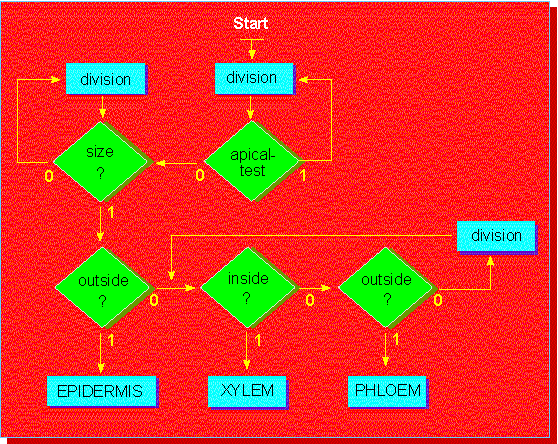
In 1965, J. BONNER of the California Institute of Technology at Pasadena presented a model that explains how genetically determined co-ordination points for the development of specialized cells develop from a single cell. It may seem rather hypothetical, but it is a useful working hypothesis that prompts to search for the demanded regulators.

|
|