
In our discussion of photosynthesis have we thus far only regarded biochemical reactions. In the section about glycolysis and other biosynthetic pathways was the significance of single enzymes pointed out. Since quite some time know, for example, have all enzymes involved in glycolysis been purified and isolated and each step of the pathway can be analyzed under in vitro conditions. It has also been tried to isolate the complete set of components necessary for photosynthesis in order to reconstitute the whole system. But all attempts failed because the premises they were based on were wrong as we know today.
A number of problems have not been taken into consideration until now. The terms photosystem I and photosystem II, for example, have been introduced and all participating pigments have been mentioned but the following subjects remain to be discussed:
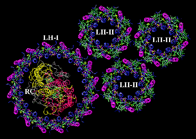
![]() How are the photosystems organized?
How are the photosystems organized?
![]() How are the pigments arranged?
How are the pigments arranged?
![]() Why does one of the chlorophyll molecules react different
than all the others?
Why does one of the chlorophyll molecules react different
than all the others?
![]() Why are action and absorption spectra not quite congruent?
Why are action and absorption spectra not quite congruent?
![]() Why reacts P 680 (chlorophyll a) different than P 700 (chlorophyll
a, too)?
Why reacts P 680 (chlorophyll a) different than P 700 (chlorophyll
a, too)?
![]() How are electron transport chain and ATP production coupled?
How are electron transport chain and ATP production coupled?
![]() How are photosystem I and II linked?
How are photosystem I and II linked?
![]() Which structural prerequisites have to exist in order for
the two systems to co-operate?
Which structural prerequisites have to exist in order for
the two systems to co-operate?
It was always accepted that each of the biochemical reactions was catalyzed by a specific enzyme and still, it took quite some time before it was realized that the chlorophyll and the other pigments are protein-bound and that they are only active as protein-chlorophyll (and protein-pigment, respectively) complexes. The isolated pigments themselves were useless for photosynthesis. The pigment-protein complex, (most) proteins of the electron transport chain as well as the catalyst of ATP synthesis (ATP synthase) are integral compounds of the photosynthesis membrane(s) (= the thylacoid membranes of algae and higher green plants, cytoplasmatic membranes of photosynthetically active bacteria and blue-green algae). The location within the membrane (at the out- or the inside, for example) and the relative arrangement of the proteins towards each other are important prerequisites of energy transformation.
This is not only true for photosynthetic reactions but also for those of the respiratory chain and for the enzymes located within the purple membrane of Halobacterium halobium (an archaebacterium using light energy for the production of ATP without an electron flow).
The requirements for energy transformation are even higher: completely intact membranes that are impermeable for protons and that enclose compartments thus maintaining a stable electrochemical gradient between inside and outside. The production of ATP is based on a directed proton dislocation paralleled by a change of the compartment's pH and of its membrane potential.
The research into the proteins essential for photosynthesis started very late. The reason is that all of them are membrane-bound which rendered it nearly impossible to isolate and characterize them with the classical methods of protein analysis.
Only after sensitive techniques like gel electrophoresis and the controlled use of detergents like sodium dodecyl sulfate (SDS) had been developed, became it possible to separate the proteins and to identify them as bands in a gel. A side product of this technique is the determination of the molecular weights of the respective polypeptide chain.
A second, independent attempt was and is the use of specific probes like fluorescence-tagged antibodies that help to find out whether a certain protein (or part of a polypeptide chain) is located at the inside or the outside of a membrane. The use of antibodies against specific proteins allows, too, to precipitate these proteins selectively since only they are able to form the extremely specific antigen - antibody complex.
Cross-linking agents render it possible to elucidate the surrounding of a molecule. And the use of specific inhibitors helps localizing their site of effect. DCMU [3-(3', 4' - dichlorphenyl) - 1,1 - dimethylurea] has since years been used to inhibit photosystem II. It has no effect on photosystem I and was therefore used by ARNON and his collaborators as an important help to study the electron transport chain that starts at photosystem I independently of that induced by photosystem II.
We know today that DCMU does not effect chlorophyll itself but a certain protein, the plastoquinone-binding protein.
A third possibility to characterize the photosynthetic membrane is the analysis of certain mutants. The single-celled alga Chlamydomonas reinhardii proved to be a good test object. Quite a range of mutants with photosynthetic defects are known. They can be grouped in four classes:
- mutants with a defect in photosystem I,
- mutants with a defect in photosystem II,
- mutants with a defect in photophosphorylation and
- mutants with a defect in the antenna complex.
It is quite striking that almost all mutants are characterized not only by the loss or change of a certain polypeptide chain but by the lack of a whole complex, for example that of PS I. It seems therefore as if the mutations would lead to pleiotropic effects. Or, expressed differently: when a polypeptide chain is changed or missing does the assembly of the other polypeptide chains not work any more. This observation shows how tight the interactions between the single polypeptide chains are and how important they are for their mutual co-operation.
A further and not less important technique is electron microscopy usually used in combination with freeze-etching.
The sequencing of membrane proteins remains difficult. And yet, the sequences of most proteins involved in photosynthesis could be determined during the last years via the sequencing of their respective genes. The most remarkable outcome of this work is that these proteins contain (just like proteins of animal or bacterial membranes, too) a large portion of alpha - helices. The lengths of the helices corresponds to the thickness of the membranes. The single helices are connected via polar and / or non-hydrophobic sequences.
Several chlorophyll-binding proteins of the photosynthetic membranes of different systematic groups (angiosperms, gymnosperms, algae, bacteria) have been isolated and characterized. The best-known are P 700-chlorophyll-a-protein 1 and the light-harvesting chlorophyll-a/b-protein 2 which have been studied in the laboratory of J. P. THORNBERGER at the University of California, Los Angeles. Both are strongly hydrophobic, integral membrane proteins. Both bind chlorophyll a but only the latter binds chlorophyll b, too. The P 700-chlorophyll-a-protein 1 contains the reaction centre (P 700) of photosystem I, i.e. one of the chlorophyll molecules is bound in a specific configuration and is located in a surrounding (due to a specific amino acid composition and the folding of the polypeptide chain) that renders it different than all other chlorophyll molecules bound by this protein, too. This structural peculiarity is the precondition for the light-induced activation and consequently for the induction of the electron flow.
The molecular weight of the polypeptide chain is 110,000 Dalton. It is able to bind 14 chlorophyll molecules. The light-harvesting-protein (light-harvesting chlorophyll-a/b-protein 2) is also very common. It is mainly associated with photosystem II but effects on photosystem I have been observed, too. Chlorophyll a and b are bound in equimolar amounts besides lutein and beta - carotene. The chlorophyll to carotenoid ratio is 3 - 7 : 1 on a molecular level.
©: Theoretical Biology Group - University of Illinois at Urbana-Champaign
ATP - synthase is the enzyme that catalyzes the synthesis of ATP. Since the production of ATP occurs not only during photosynthesis but during respiration, too, suggested the idea that the ATP production of both cases is based on similar mechanisms itself.
After having collected experience with the mitochondrial ATP synthase, did E. RACKER of the Cornell University isolate an enzyme of thylacoid membranes that resembled the respective mitochondrial enzyme very much. In the electron microscope did it look like a stemmed knob. The knob was termed CF1 and the stem CF0 (F1 and F0 respectively in the mitochondrial enzyme). CF0 (or F0) is a membrane anchor. While the F1 - F0 complex is localized in the inner mitochondrial membrane and the knob is directed towards the mitochondrial matrix are the respective molecular parts of the CF0 - CF1 complex found at the outside of the thylacoid membranes. In both cases consists the ATP synthase out of several different polypeptide chains, it is an enzyme complex. The phosphorylation of ADP works only, if the ATP synthase is a component of an intact, proton-permeable membrane. It has to separate two compartments (the inner part of the vesicle and the surrounding).