

|
|
Chromosomes are structures within a cell which contain the genetic information that is passed on from one generation to the next.
In prokaryotes, there is usually a large circular chromosome. In addition, there may be several smaller circular chromosomes called plasmids. None of these is enclosed in a membrane, they merely drift around within the cytoplasm of the prokaryote cell.
In eukaryotes, most chromosomes are located in the cell nucleus and are composed of protein and DNA. Smaller chromosomes exist in mitochondria and chloroplasts - these tend to regulate activity solely within the organelle in which they occur.
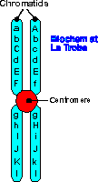
| Chromosomes only become visible through a microscope just prior to cell division. Before they are able to be seen, the DNA has been duplicated and coils tightly around proteins. The two strands of DNA so formed are called chromatids and are still joined by a protein disk at a point called the centromere. |  |
Human cells other than gametes (= somatic cells) contain 46 chromosomes. This represents 22 pairs of autosomes and a pair of so called sex chromosomes. When a cell has a set of paired chromosomes, as in a somatic cell, it is described as diploid.
The chromosomes in each autosome pair (= homologous chromosomes) look very similar in length, position of the centromere and staining reactions. And when the genetic information on a pair is examined it is found that the genes match up perfectly although the proteins for which they code may have important differences.
The sex chromosomes may or may not be homologous. In a human female, each somatic cell has two copies of the X chromosome as well as the 22 autosome pairs. The human male has 22 pairs of autosomes and one X chromosome and one Y chromosome. The Y chromosome is physically smaller than an X chromosome and only some of the genetic information on an X chromosome has matching information on the Y chromosome.
Other species have different numbers of chromosome pairs in their somatic cells. It is this difference in size and arrangement of chromosomes which, at the molecular level, separates one species from the other.
It is possible to stain the chromosomes of a dividing cell and view them under a microscope. If the image is then photographed and enlarged, the individual pictures of the chromosomes can be cut up and sorted. This is known as a karyotype.
Examination of an individual's karyotype may reveal information about gender or the presence/absence of the correct number of chromosomes. For this reason karyotype analysis is sometimes performed on cells from unborn babies.
A gene is a section of a chromosome. A gene carries coded information within a sequence of chemicals. When appropriate, a section of this information is decoded and used to provide the recipe for protein synthesis within the cell. The proteins synthesised may affect the way the cell interacts with its environment. This, in turn, may affect the way the whole organism responds to its environment.
Since the genes directly control the functioning of cells, it is hardly surprising that (almost) all cells contain chromosomes. When cell reproduction occurs (as in growth or tissue repair) it is therefore necessary to reproduce the chromosomes in such a way that each new cell has its full quota of the DNA needed to ensure the cell's survival.
The process of mitosis ensures that each new (daughter) cell in the organism's body inherits the correct amount of DNA. This is described in more detail on the page "New proteins, Cells and Individuals".
Similarly, new individuals need a supply of DNA in order to exist. If these individuals are generated by asexual reproduction, they are provided with a copy of the same chromosomes, and genes, as the parent from which they arise. Individuals which are the product of sexual reproduction, however, gain half of their DNA from each parent. A different type of cell reproduction is called for in preparation for sexual reproduction if the cells of the offspring are not to be overloaded with too many chromosomes.
The process of meiosis results in gametes with half as many chromosomes as a normal body cell (a somatic cell). Gametes from two parents unite in sexual reproduction to form a zygote with the correct amount of DNA. This is described in more detail on the page"New proteins, Cells and Individuals".
Deoxy-ribo-nucleic acid (DNA) is the chemical which carries the genetic code.
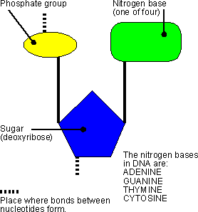
DNA is composed of sub-units called nucleotides (see diagram at right), which themselves are composed of sub-units: a sugar molecule (deoxyribose), a phosphate group, and one of four possible nitrogen bases. The nucleotides in a single strand of DNA are linked together via chemical bonds between the phosphate group of one nucleotide and the ribose of the next. In this way, a backbone of -ribose-phosphate-ribose-phosphate-ribose-phosphate- is formed with the bases attached to each ribose. |
 |
It was discovered in 1949 that there is always the same amount of adenine and thymine in DNA and similarly, there is always the same amount of cytosine as guanine. From this observation, in 1953, Watson and Crick (and others) deduced the structure of DNA to be two single strands, wound around each other, and linked together with weak chemical bonds between the bases on each strand. The specific base pairing was found to always occur between adenine:thymine and guanine:cytosine, thus accounting for the constant ratios observed earlier. The arrangement of two strands of DNA, linked by the base pairs and twisted together, is described as a double helix.
Because of the special relationship between the bases of one strand of DNA with the bases on the opposite strand, the two chains of the double helix are described as complementary to each other.
There is almost certainly a daigram of DNA in your textbook, but if you can be bothered waiting for a rather large picture from the USA you can click here. Use your back button to come back here from the DNA picture.
When cells prepare to divide, either for growth or for repair, the chromosomes become visible as duplicated threads, the chromatids. For chromatids to form, the DNA of the original chromosomes needs to be reproduced so that each new cell will ultimately possess the correct genetic information to function appropriately.
 |
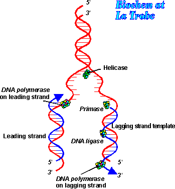
DNA replication occurs when the complementary strands of DNA break apart and unwind. This is accomplished with the help of enzymes called helicases. Additional enzymes and proteins attach to the individual strands, holding them apart and preventing them from coiling upon themselves. The point at which the double helix separates is called the replication fork, because of the shape of the molecule. At this site enzymes called DNA polymerases move along each of the separated DNA strands, adding nucleotides to the exposed bases according to the base pairing rules. The ribose-phosphate bonds form between the new nucleotides to hold the new strand together. The process continues until the original double helix is completely unwound and two new double helices have been formed. Each new double helix is composed of one old DNA strand and one new strand. This is described as semi-conservative replication. |
During replication, there are occasionally errors made in the insertion of the complementary bases in the new DNA strand. Usually these are repaired by special DNA polymerases, but a few remain (increasing in frequency with the increasing age of the organism). These errors in the DNA may or may not have an effect on the functioning of the cell lines in which they then appear. Errors or alterations in the base sequence of DNA are called mutations.
One of the most important discoveries in Biology was the realisation that genes are responsible for the production of proteins. George Beadle and Edward Tatum, working at the California Institute of Technology in the 1940s are credited with this discovery, although, of course, they were working on extending ideas that had been published earlier by other scientists. Then it was Alfred Hershey and Martha Chase who, in 1952, published the results of their experiments that convinced scientists that DNA is indeed the carrier of hereditary information.
So how does the DNA carry the 'recipe' for all the proteins, structural and functional, that an organism needs? The answer is in the four bases, Adenine, Thymine, Cytosine and Guanine (learn how to spell these!). These four bases on the DNA chain in the double helix can be thought of as 'letters' which are used to build the total possible range of three letter 'words' - it's a simple maths exercise to realise that this gives a possible 'vocabulary' of 64 (=4x4x4) words.
Since there are only 20 amino acids that are used, in various combinations, to make proteins, it is possible to give each amino acid a DNA code 'word'. For example, the DNA triplet code CGA will lead to the insertion of the amino acid alanine into a protein. But there are still 'words' left over to have duplicates for many of the amino acids (CGG, CGT and CGC also code for alanine) and 'special words' to tell where a gene stops (ATT does this).
The term for the DNA bases in their groups of three is codons, and it is these codons, grouped together as genes, which hold the information for the accurate production of all the cells' proteins. That is the subject of the next page at this site.