
![]()
![]()
![]()
![]()
![]()
![]()
![]()
| Topics covered on this page are:
|
 |
Exactly how does the DNA in a portion of a chromosome (i.e. the DNA in a gene) determine an individual's characteristics? The answer is, in fact, to be found in the proteins which are coded for by the DNA.
Different proteins do different things; structural proteins determine the organisms physical form, functional proteins determine how the cells, tissues, organs and indeed the whole organism survives in its environment, and regulatory proteins act as switches where appropriate to direct metabolic responses as needed.
How, then, does DNA determine the nature of a protein?
Transcription is the formation of RNA from a DNA template. The RNA formed may be rRNA (ribosomal RNA), tRNA (transfer RNA) or mRNA (messenger RNA). The functions of rRNA and tRNA are discussed elsewhere on this page. At present, we will concentrate on the formation of mRNA by the process of transcription.
A flow chart will summarise the steps involved, but students should read this in conjunction with a series of good quality diagrams which illustrate the process.
Inside the nucleus of a eukaryotic cell, an enzyme, RNA polymerase, binds to a specific sequence of bases on the DNA of the gene to be expressed. This starting sequence is the promotor.
The DNA begins to unwind and the strands begin to separate.
| BASE ON DNA SENSE STRAND | COMPLEMENTARY BASE ON mRNA |
|---|---|
| |
|
Transcription (literally "rewriting") continues until the RNA polymerase reaches a "stop" message on the DNA (a terminator).
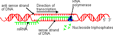
Before leaving the nucleus, the mRNA is modified so that only the sections of it necessary for the "recipe" of the required protein are retained. This is necessary because the DNA of a eukaryotic gene seems to contain large lengths of "junk" information, called introns, which interrupt the sections which will ultimately be expressed, the exons. (In DNA from prokaryotes, this is not the case. There is very little, if any, intron material in bacterial genes.)
Both introns and exons are transcribed onto mRNA, but enzymes cut out the introns and rejoin the exons into the final molecule of mRNA which leaves the nucleus. The mRNA leaves the nucleus, via nucleopores, and travels in the cytoplasm to a ribosome.
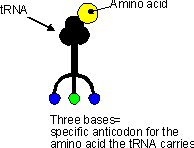
It is at the ribosome that the base sequence code of the mRNA is translated into the string of amino acids which will ultimately form a protein. This translation is possible because the sequence of the bases on the mRNA is "read" in groups of three. Each group of three bases on the mRNA is called a codon. Each codon is specific for the insertion of one amino acid into the growing protein on the ribosome. This is described on the page of this site titled "Chromosomes, Genes and DNA".
Ribosomes are made up of two subunits, a large one and a smaller one. Each ribosome consists of ribosomal RNA (rRNA) and proteins (about 70 different types). Ribosomes are either found attached to the cell's internal membrane system (rough endoplasmic reticulum) or free in the cytoplasm.
Transfer RNA (tRNA) is also involved in the synthesis of proteins. This substance is a single strand of RNA that loops back on itself. At one of the free ends, there may be an amino acid attached to the tRNA. At the central loop, there are three bases (the anticodon) exposed. There are different tRNAs for each of the 20 amino acids. The sequence of the three bases in the anticodon determines which amino acid a particular tRNA will carry. |
|

|
This tRNA carries the amino acid methionine. and peptide bonds forming between the amino acids. |
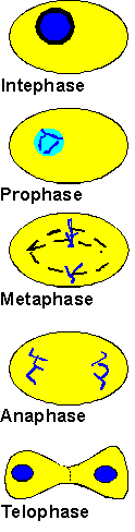
Mitosis is the process by which the nucleus of a cell is divided into two nuclei, each with the same number and kinds of chromosomes as the parent cell. Mitosis is the process by which all of an organism's body cells (the somatic cells) are reproduced.
The process of mitosis is divided into phases in which particular events take place. The acronym IPMAT can be used to remember the phases in order.
|
|
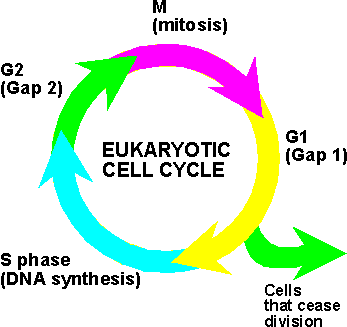
The cell cycle is the period from the end of one mitosis to the end of the next. During a cell cycle, a cell grows (the G1 gap phase in the diagram), prepares for division (the S phase), continues to function for a while (G2 phase) and divides to form two daughter cells (M phase), each of which begins the cycle for itself. The time to complete one cell cycle varies considerably with different cells. Most human nerves and muscle cells do not divide once formed, but the cells lining the digestive tract may pass through a complete cell cycle every six hours. |
 |
Meiosis is the process that produces haploid gametes from diploid cells. This process reduces the number of chromosomes in the gametes to half of that in the normal somatic cells. During meiosis the homologous chromosomes which exist in diploid cells are separated. In humans this means that sperm and ova contain 23 chromosomes (the haploid number) but somatic cells contain 46 chromosomes (the diploid number). The desirability of this becomes apparent when fertilization is considered.
The phases of meiosis can be remembered as IPMAT-PMAT. Whilst the names of the phases of meiosis seem to be the same as those of mitosis, there are some very important differences in the details of meiosis compared with mitosis. When reading the stages which follow, students are advised to have a detailed diagram of meiosis available as an additional reference.
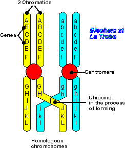
| At the start of meiosis, during prophase I, the pairing of the homologous chromosomes means that the four chromatid strands can become intertwined. At this time the strands may be exposed to pressure at points of overlap (called chiasma - plural chiasmata) and breaks may occur. Enzymes quickly repair these breaks, but often the "wrong" strands are rejoined, leading to new combinations of genetic information in the repaired chromatids. This process is known as crossing over and it can result in either more or less desirable combinations of genes in the gametes which are eventually formed and contain the new gene combinations. This, in turn, leads to individuals with new characteristics which may make them better or worse suited to survival. The resulting variation amongst individuals in a population is a major contributor to the explanation of Natural Selection (more about that later). |
 |
The genotype of an individual refers to that individual's genetic makeup. It is the total of all the genes that person possess on his/her chromosomes. Often, in discussing inheritance, we consider only a few of the individual's genes and then the term genotype refers to the particular combination of these genes of interest that the individual possesses.
Because somatic cells have two copies of each chromosome (the homologous pairs), each gene is represented twice in each cell. Normal homologous chromosomes always have the same gene at the same position (the locus -plural loci). But mutations may have, over many generations, caused slight differences in the base sequences of the genes in different individuals (and in the gene products), so that the gene may exist in two or more different forms. These different forms of a gene are termed alleles.
The alleles of any particular gene are often represented by letters. If there are only two possible alleles, capital and lower-case letters are used. So for a gene with two alleles, we could label the alleles A and a. If the individual has two alleles the same, either genotype AA or aa, this is described as being homozygous. An individual with different alleles has genotype Aa and is heterozygous. (Some texts use the term hybrid as a synonym for heterozygous, but this can lead to confusion and should be avoided.)
Some alleles are found to be dominant over their alternative allele. This means that if an individual has either one (genotype Aa) or two (AA) copies of the allele its effects will always be seen. A dominant allele is always assigned a capital letter, hence it is A in the example given. The alternative allele (a), is said to be recessive. The effects of the recessive allele are only observed when no dominant allele is present, that is in an individual who is homozygous for the recessive allele.
The observable effects of the alleles an individual possesses are called the phenotype of that individual.
Depending on the nature of the genes in question, the phenotype may be structural (some particular visible characteristic, eg fur colour), functional (something to do with the organism's metabolism, eg ability to digest lactose) or behavioural characteristics (the ability to do something, eg make sexy movements which attract many mates). Clearly, different methods need to be used to 'observe' the different phenotypes described above, but the important point is that the differences can, with appropriate methods, be observed.
In the simplest cases, only one gene, and its alleles, controls a particular characteristic (or trait). An example of this is the famous tall and short pea plants studied by Mendel. But in many cases, a number of genes (and all of their possible alleles) have an effect on a particular trait. This is polygenic inheritance and is responsible for the continuous variation seen in some traits - for example human height. This is discussed further on the page Real-world Genetics.
Whilst the genotype of an individual determines what that individual can become, it is not the only factor in determining the individual's phenotype. For example, with inadequate nutrition and exercise, a person with the genetic potential to be an Olympic heavyweight weightlifting champion may end up as a proverbial "ninety pound weakling".
Similarly, when an apparently healthy population of individuals moves from one location to another location with markedly different conditions it may be found that some individuals possess previously unknown abilities to survive in the new conditions whilst others rapidly deteriorate.
This relationship between an individual's genotype, the environment in which he/she/it lives and the observed phenotype is often summarised in the following "equation":
Phenotype = Genotype + Environment |
A mutation is a change in the genetic material in a cell. That is, the sequence of bases in a part of the cell's DNA is altered in some way. Since the processes of duplicating genetic information and passing it on to either the next generation of cells or the next generation of individuals are complex, it is hardly surprising that mistakes sometimes occur. These mistakes lead to the changes which are mutations.
Not all mutations are harmful. Many mutations have no effect or cause minor harmless effects in the organism. Occassionally a mutation may be beneficial to the organism. Sometimes, however, mutations are harmful or even lethal. The effect of a mutation is entirely dependent on the amount and location of the DNA which it alters.
Chromosomal mutations involve segments of chromosomes, whole chromosomes, and even entire sets of chromosomes.
There are four types of chromosomal mutations:Nondisjunction is the cause of chromosomal mutations which involve whole chromosomes or sets of chromosomes. This is the failure of homologous chromosomes to separate properly during meiosis (see above). The resulting gametes will have more or fewer chromosomes than they normally would.
Nondisjunction of human chromosomes can lead to a number of disorders. Sometimes it is fatal, depending on the particular chromosomes involved.
When all the homologous chromosomes fail to separate during meiosis the gametes which are formed are not haploid. Depending on the other gamete involved in fertilization, the resulting offspring may be triploid (3N) or tetraploid (4N). This condition is known as polyploidy and is almost always lethal in animals. Polyploid plants, however, are often larger and hardier than normal plants.
Gene mutations involve individual genes.
The smallest gene mutation is a point mutation where only one base is affected. If this is just a change to a different base, the effect may be minimal, although if the change causes a change in the amino acid sequence of a polypeptide the effect may be major.
However, if a single base is added to or deleted from a gene, the whole sequence of bases is altered and so the codons become changed. This is a frameshift mutation and it can completely change the polypeptide product of the gene. The extent of the change in the polypeptide product depends upon where, in the gene, the frameshift mutation occurs. The consequences of this, in terms of the functioning of the organism can be disastrous.
A somatic mutation is one which occurs during mitosis in an individual's body cell. This type of mutation will be passed on to all cells in the cell line in which it arose. If there are still plenty of normal cells surrounding the mutated cell/cells, the normal cells may continue to provide enough normal gene product that the affected cells do not influence the functioning of the organism.
Mutations also occur in mitochondrial DNA. These are similar to somatic mutations since mitochondrial DNA (mtDNA) reproduces by a mechanism similar to mitosis. Since this mechanism does not allow for 'crossing over' and the mixing of genes seen in meiosis, changes accumulate much more slowly in mtDNA than in chromosomal DNA. Also, since mitochondria are inherited along the maternal side of any population (think about the structure of the ovum and the sperm and the details of fertilisation to explain this), this too slows the rate of accumulation of mtDNA mutations in a population. Mitochondrial DNA mutations are of interest to scientists in establishing 'relatedness' between individuals, especially in attempting to determine evolutionary links between and within species.
Mutations which affect the ability of cells to regulate their own growth and reproduction may cause cancer. In this case tumours may form and the normal functioning of tissues may be so severely impaired that death occurs.
A mutagen is a substance or agent which increases the frequency of mutations in organisms. Many different chemicals (such as nicotine) are known to be mutagens as are many forms of radiation (eg X-Rays). A mutagen which causes cancer is known as a carcinogen.