 X-ray structure of flock house virus, colored by its proteins.
 X-ray structure of rhinovirus colored by its proteins.
 X-ray structure of rhinovirus radially depth cued.
 X-ray structure of rhinovirus 16, shown with 2 pentamers
removed so as to see the VP4 protein on the interior.
X-ray structure of rhinovirus 16, shown with 2 pentamers
removed so as to see the VP4 protein on the interior.
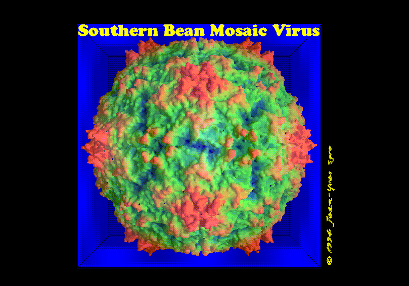 Molecular surface of Southern Bean Mosaic Virus, radially depth cued,
from x-ray data.
Molecular surface of Southern Bean Mosaic Virus, radially depth cued,
from x-ray data.
 Molecular surface of Southern Bean Mosaic Virus, radially
depth cued in blue, from x-ray data.
Molecular surface of Southern Bean Mosaic Virus, radially
depth cued in blue, from x-ray data.
 Molecular surface of Southern Bean Mosaic Virus, radially depth cued,
from x-ray data.
View along the icosahedral 2-fold axis.
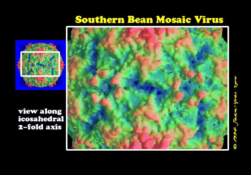
Molecular surface of Southern Bean Mosaic Virus, radially depth cued,
from x-ray data.
View along the icosahedral 2-fold axis.
 Molecular surface of Southern Bean Mosaic Virus, radially depth cued in
blue, from x-ray data. View along the
icosahedral 2-fold axis.
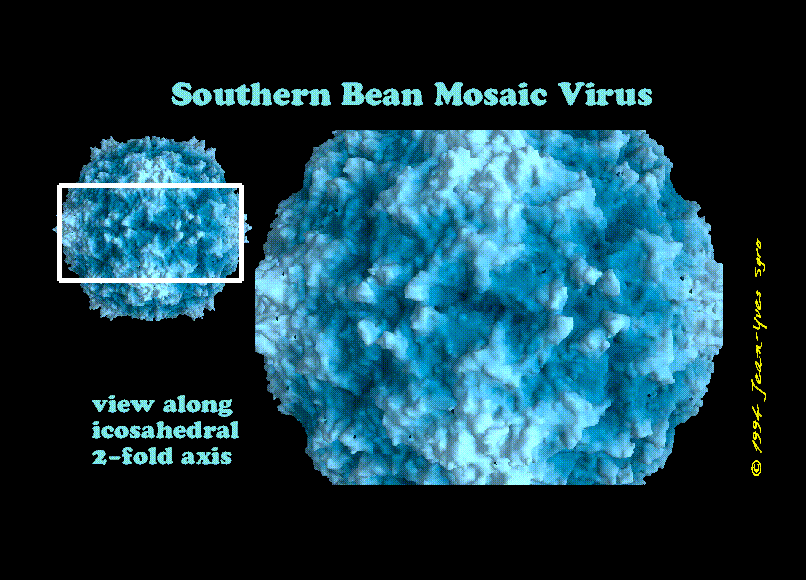
Molecular surface of Southern Bean Mosaic Virus, radially depth cued in
blue, from x-ray data. View along the
icosahedral 2-fold axis.
 Go up to the
Scientific Visualization at the IMV page
Go up to the
Scientific Visualization at the IMV page
 Go to the
top page
Go to the
top page
© 1994 Jean-Yves Sgro. Institute for Molecular Virology/ jsgro@facstaff.wisc.edu