Computer Visualizations of Viruses - Part 2
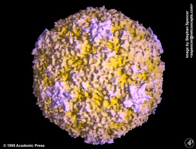
 Rhinovirus 14, as solved by cryo-electron microscopy and image reconsruction courtesy of Tim Baker. Purdue University
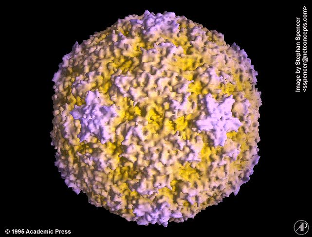
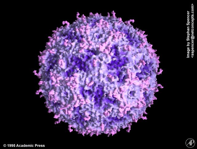
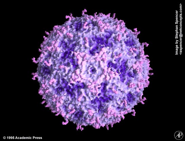
 Rhinovirus 14, color coded by protein, as solved by X-ray crystallography
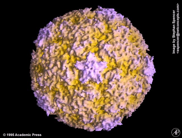
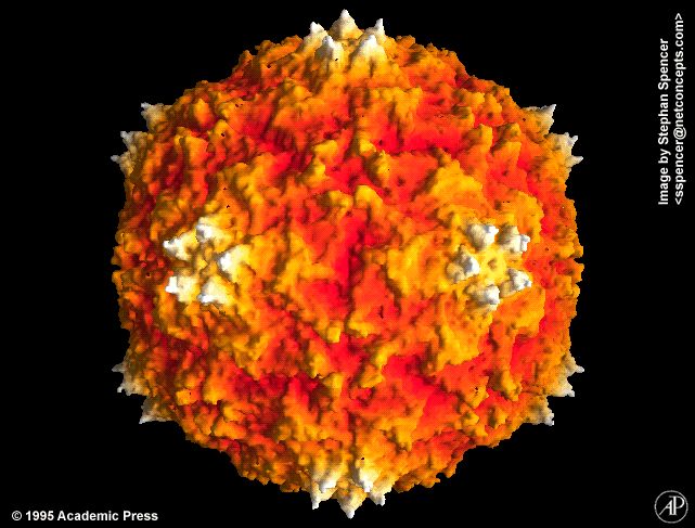
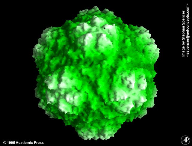
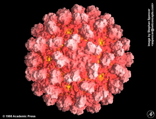
 Molecular surface of Rhinovirus 14, radially depth cued,
as solved by X-ray crystallography
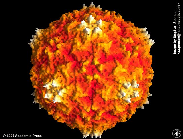
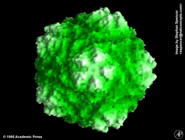
Molecular surface of Rhinovirus 14, radially depth cued,
as solved by X-ray crystallography
View along the 2-fold axis
More views along the
2-fold axis,
3-fold axis,
5-fold axis *
180 degree rotation (1.6MB Quicktime movie) *
Rhinovirus shown on 20/20 on abc TV.
(quicktime and animated Gifs).
 Rhinovirus 14, radially depth cued with antigenic sites highlighted, as solved by X-ray crystallography
 Rhinovirus 14 complexed with neutralizing antibodies, as solved by cryo-electron microscopy and image reconstruction courtesy of Tim Baker. Purdue University
 Rhinovirus 14 complexed with the ICAM-1 receptor, as solved by cryo-electron microscopy and image reconstruction courtesy of Tim Baker. Purdue University
 Rhinovirus 16 with view of the interior, as solved by X-ray crystallography
Ross River Virus
Electron density isosurface of Ross River Virus, radially depth cued,
as solved by cryo-electron microscopy and image reconstruction. (Structure solved by
Tim Baker of Purdue University.)
Views along the
2-fold axis,
3-fold axis,
5-fold axis *
180 degree rotation (1.5MB Quicktime movie) *
Southern Bean Mosaic Virus
 Molecular surface of Southern Bean Mosaic Virus, radially depth cued,
as solved by X-ray crystallography
Molecular surface of Southern Bean Mosaic Virus, radially depth cued,
as solved by X-ray crystallography
View along the 2-fold axis
More views along the
2-fold axis,
3-fold axis,
5-fold axis *
180 degree rotation (1.6MB Quicktime Movie) *
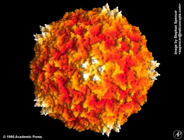
 Molecular surface of Southern Bean Mosaic Virus, radially depth cued,
as solved by X-ray crystallography
Molecular surface of Southern Bean Mosaic Virus, radially depth cued,
as solved by X-ray crystallography
 Close up view of the molecular surface of Southern Bean Mosaic Virus, radially depth cued,
as solved by X-ray crystallography
Close up view of the molecular surface of Southern Bean Mosaic Virus, radially depth cued,
as solved by X-ray crystallography
 Close up view of the molecular surface of Southern Bean Mosaic Virus, radially depth cued in
blue, as solved by X-ray crystallography
Close up view of the molecular surface of Southern Bean Mosaic Virus, radially depth cued in
blue, as solved by X-ray crystallography
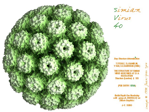
Simian Virus 40
 Molecular surface of Simian Virus, radially depth cued,
as solved by X-ray crystallography
Molecular surface of Simian Virus, radially depth cued,
as solved by X-ray crystallography
Satellite Tobacco Necrosis Virus
Molecular surface of Satellite Tobacco Necrosis Virus,
radially depth cued, as solved by X-ray crystallography
Views along the
2-fold axis,
3-fold axis,
5-fold axis *
180 degree rotation (1.3 MB Quicktime movie) *
Theiler's Murine Encephalomyelitis Virus
Molecular surface of Theiler's Murine Encelphalomyelitis Virus,
radially depth cued, as solved by X-ray crystallography
Views along the
2-fold axis,
3-fold axis,
5-fold axis *
180 degree rotation (1.4 MB Quicktime movie) *
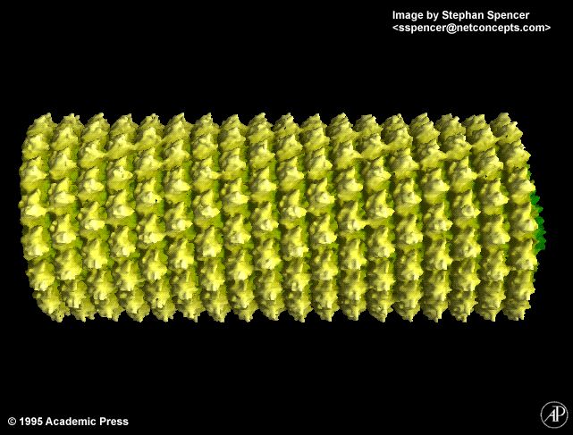
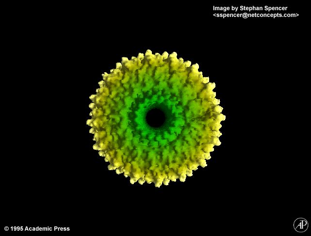
Tobacco Mosaic Virus
Molecular surface of Tobacco Mosaic Virus, radially depth cued,
as solved by X-ray crystallography
Views
along the helical axis,
down the helical axis *
180 degree rotation (1.1 MB Quicktime movie) *
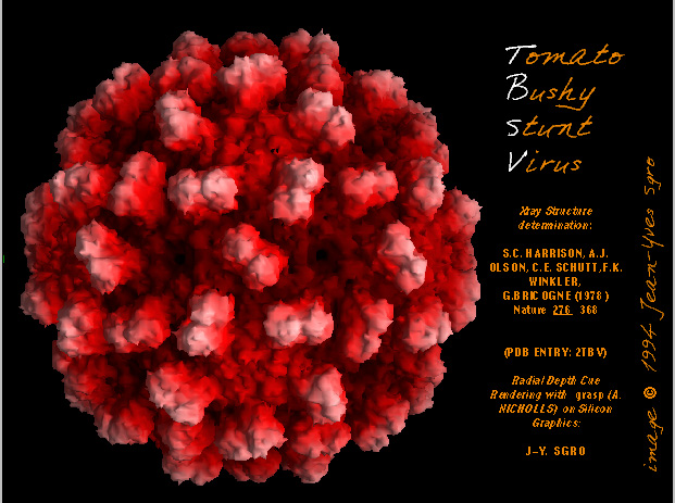
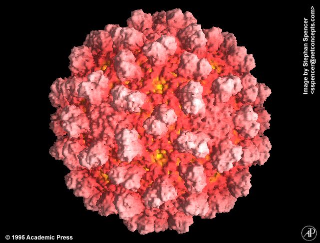
Tomato Bushy Stunt Virus
 Molecular surface of Tomato Bushy Stunt Virus, radially depth cued,
as solved by X-ray crystallography
Molecular surface of Tomato Bushy Stunt Virus, radially depth cued,
as solved by X-ray crystallography
View along the 2-fold axis
More views along the
2-fold axis,
3-fold axis,
5-fold axis *
180 degree rotation (1.6MB Quicktime Movie) *
Turnip Yellow Mosaic Virus
 Molecular surface of Turnip Yellow Mosaic Virus,
radially depth cued, as solved by X-ray crystallography
Molecular surface of Turnip Yellow Mosaic Virus,
radially depth cued, as solved by X-ray crystallography
Simulations
 Simulation of a virus binding to a host cell
receptor
Simulation of a virus binding to a host cell
receptor
 Morph animation of mammalian reovirus virion to ISVP transition
 Morph animation of mammalian reovirus ISVP to core transition at a vertex
 Mammalian reovirus ISVP with simulated flexible spikes
References
- Spencer SM, Sgro JY, Dryden KA, Baker TS, Nibert ML
IRIS explorer software for radial-depth cueing reovirus
particles and other macromolecular structures determined
by cryoelectron microscopy and image reconstruction.
J Struct Biol 1997 Oct; 120(1):11-21
- Nicholls A, Sharp KA, Honig B
Protein folding and association: insights from the interfacial
and thermodynamic properties of hydrocarbons.
Proteins 1991;11(4):281-296
* Visualizations designated with an asterisk (*) are
reproduced with permission from the Encyclopedia of
Virology Plus CD-ROM. (Edited by Robert G.
Webster and Allan Granoff.
© 1995 Academic Press Ltd
All Rights Reserved.
Go on to
Part 1
Go back to the
Visualizations of Viruses page
Content: ©1994-1999 Stephan Spencer &
Jean-Yves Sgro
Design: ©1997
Internet Concepts, LLC
Web Design by


